The Aorta is the largest artery (arterial trunk) of the body which carries the oxygenated blood from the left ventricle and supplies it to all the parts of the body.
Parts Of The Aorta
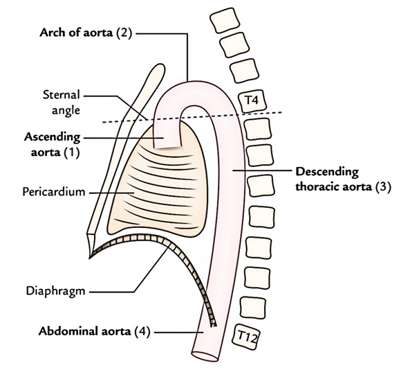
For the convenience of description, the aorta is splited into the following 4 parts:
- Ascending aorta.
- Arch of aorta.
- Descending thoracic aorta.
- Abdominal aorta.
The first 3 parts are confined to the thoracic cavity and together create the thoracic aorta.
Ascending Aorta Origin And Course
A. Ascending aorta originates from the upper end of the left ventricle (i.e., aortic vestibule) and continues as an arch of aorta at the sternal angle.
B. It’s about 5 cm long and its diameter is about 3 cm. It’s completely enclosed in the pericardium. It begins behind the left half of the sternum in the level of the lower border of left 3rd costal cartilage, runs upwards, forwards and to the right to continue as the arch of aorta at the level of sternal angle.
Aortic Sinuses (Sinuses Of The Valsalva)
The root of aorta presents 3 dilatations termed aortic sinuses of Valsalva. These dilatations are just above the cusps of the aortic valve. These positions are: anterior, left posterior, and right posterior.
A. Anterior aortic sinus supplies origin to the right coronary artery, therefore it’s also named right coronary sinus.
B. Left posterior aortic sinus supplies origin to the left coronary artery, therefore it’s also referred to as left coronary sinus.
C. Right posterior aortic sinus is called non-coronary sinus.
Aortic bulb is a bulge in the right wall of the ascending aorta at its union with all the arch of the aorta.
Relations
Anterior
From below upwards these are as follows:
A. Infundibulum of right ventricle.
B. Pulmonary trunk.
C. Pericardium.
Posterior
From before backwards and to right these are as follows:
A. Transverse sinus of pericardium.
B. Right pulmonary artery.
C. Right principal bronchus.
To The Right:
A. Right atrium.
To the left:
A. Left atrium.
B. Pulmonary trunk.
Branches
A. Right coronary artery from anterior aortic sinus.
B. Left coronary artery from left posterior aortic sinus.
Development
The ascending aorta develops from the truncus arteriosus after its partition by the spiral septum.
Clinical Significance
Aneurysm of ascending aorta: It takes place at the bulb of the ascending aorta. The bulb of aorta is a dilatation in the right wall of ascending aorta that is subjected to constant thrust of the forceful blood current ejected from the left ventricle. It might compress the right atrium, SVC or right principal bronchus. Its rupture (a serious complication) leads to accumulation of blood in the pericardial cavity (hemopericardium).

 (60 votes, average: 4.37 out of 5)