Azygos is a term which means single that means without a companion. Azygos, hemiazygos, and accessory hemiazygos veins together compose the azygos system. The front of thoracic part of vertebral column is where these veins are located and play a significant role in the venous drainage of the thorax.
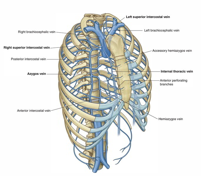
Azygos Vein
The azygos vein is present only on the right side in the upper part of the posterior abdominal wall and the posterior mediastinum. It attaches the inferior vena cava together with the superior vena cava. It comprises of valves and might appear tortuous.
The functions of azygos vein are as follows:
- It drains venous blood from the thoracic wall and upper lumbar region.
- It creates an essential collateral channel attaching the superior vena cava and inferior vena cava.
Formation
The formation of azygos vein is variable. It’s created in one of the following ways:
- Created by the union of right subcostal and right ascending lumbar vein in the level of T12 vertebra (common).
- Originates from the posterior aspect of the inferior vena cava (IVC) near the renal veins.
- As a continuation of right subcostal vein.
- Occasionally, it might originate from right renal or right first lumbar vein.
Course And Termination
The azygos vein after formation ascends up and leaves the abdomen by going through the aortic opening of the diaphragm and enters the posterior mediastinum. There it ascends vertically being located in front of vertebral column up to the level of T4 vertebra, where it arches forwards above the hilum of the right lung to terminate in the superior vena cava in the level of the 2nd costal cartilage.
Relations
Anterior: Esophagus (right edge).
Posterior:
- Lower 8th thoracic vertebrae.
- Right posterior intercostal arteries.
To the right:
- Right lung and pleura.
- Greater splanchnic nerves.
To the left:
- Thoracic duct.
- Descending thoracic aorta.
- Esophagus (right border).
The arch of azygos vein is related below to the root of right lung, on right side to the right lung and pleura, and left side to the right border of esophagus, trachea, and right vagus nerve.
Tributaries
The tributaries of the azygos vein are as follows:
- Lower 7th right posterior intercostal veins with the exception of first.
- Right superior intercostal vein (created by union of 2nd, 3rd, and 4th right posterior intercostal veins).
- Hemiazygos vein (at the level of T7 or T8 vertebra).
- Accessory hemiazygos vein (at the level of T8 or T9 vertebra).
- Right subcostal vein.
- Right bronchial vein.
- Right ascending lumbar vein.
- Esophageal veins with the exception of those at its lower end.
- Mediastinal veins.
- Pericardial veins.
Clinical Significance
In case of obstruction of SVC, it acts as the main collateral channel to shunt the blood from the upper half of the body to IVC .
Hemiazygos Vein
The hemiazygos vein (syn. inferior hemiazygos vein) is located on the left side only and corresponds to the lower part of the azygos vein (i.e., mirror image of the lower part of the azygos vein).
Formation
The hemiazygos vein created on the left, quite similar to the azygos vein, by the union of left ascending lumbar vein and left subcostal vein. It might originate from the posterior surface of the left renal vein.
Course And Termination
It pierces the left crus of the diaphragm and ascends vertically in front of the left side of the vertebral column up to the level of T8 vertebra. At T8 vertebra it turns to the right and crosses in front of the vertebral column posterior to the aorta, esophagus and thoracic duct to terminate in the azygos vein.
Tributaries
The tributaries of the hemiazygos veins are as follows:
- Lower 3 (9th-11th) left posterior intercostal veins.
- Left subcostal vein.
- Left ascending lumbar vein.
- Small esophageal and mediastinal veins.
Accessory Hemiazygos Vein
The accessory hemiazygos vein (syn. superior hemiazygos vein) is located on the left side only and corresponds to the upper part of the azygos vein (i.e., mirror image of the upper part of the azygos vein).
Course And Termination
The accessory hemiazygos vein begins at the medial end of left 4th or 5th intercostal space and descends to the left side of the vertebral column. At the level of T8 vertebra, it turns to the right enters in front of the vertebral column posterior to the aorta, esophagus and thoracic duct to terminate in the azygos vein.
Sometimes the terminal parts of hemiazygos and accessory hemiazygos veins join together to create a common trunk which crosses across the vertebral column to open into the azygos vein.
Tributaries
The following are the tributaries of accessory hemiazygos vein:
- Fifth to eighth (5th-8th) left posterior intercostal veins.
- Left bronchial veins (sometimes).

 (82 votes, average: 4.61 out of 5)