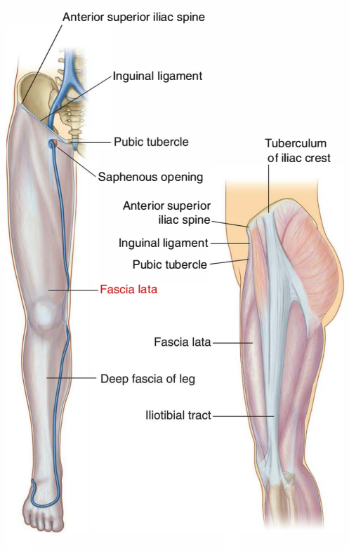
The Fascia Lata is a tough fibrous sheath that envelops the whole of the thigh like a sleeve. It is named from its great extent. “Latus” give the superlative “Latissimus” which means broadest or widest.
Attachments
Superiorly, it is attached to the boundary line between the lower limb and the pelvis.
Anteriorly, it is attached to the inguinal ligament; laterally to the iliac crest and Posteriorly, through the gluteal fascia to the sacrum, coccyx and sacrotuberous ligament.
Medially, to the pubis, the pubic arch and the ischial tuberosity and Inferiorly, on the front and sides of the knee, the fascia lata is attached to subcutaneous bony prominences and the capsule of the knee joint.
Posteriorly, the fascia lata forms the strong popliteal fascia which is continuous below with the fascia of the back of the leg.
Function
- The fascia lata surrounds the tensor fasciae latae muscle. This encircling of the muscle allows the muscles to be bound together tightly.
- The fascia lata is a fibrous sheath that encircles the thigh subcutaneously.
Modifications of Deep Fascia Lata
The deep fascia of the thigh or fascia lata presents two modifications iliotibial tract and saphenous opening.
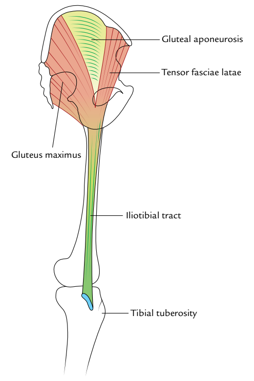
Iliotibial Tract
The fascia lata is thickened laterally where it forms a 5 cm wide band called the iliotibial tract. Superiorly, the tract splits into two layers. The superficial lamina is attached to the tubercle of the iliac crest, and the deep lamina to the capsule of the hip joint.
Inferiorly. the tract is attached to a smooth area on the anterior surface of the lateral condyle of the tibia.
The importance of the iliotibial tract is as follows:
- Two important muscles are inserted into its upper part, between the superficial and deep laminae. These are the three-fourths part of the gluteus maximus; and the tensor fasciae latae.
- The iliotibial tract stabilizes the knee both in extension and in partial flexion: and is therefore used constantly during walking and running. In leaning forwards with slightly flexed knees, the tract is the main support of the knee against gravity.
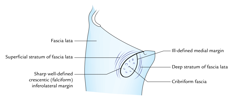
Saphenous Opening of Fascia Lata
This is an oval opening in the fascia lata. The centre of the opening is 4 cm below and 4 cm lateral to the pubic tubercle. It is about 2.5 cm long and 2 cm broad with its long axis directed downwards and laterally.
The opening has a sharp crescentic lateral margin or falciform margin which lies in front of the femoral sheath. The medial margin of the opening lies at a deeper level. It is formed by the fascia overlying the pectineus. The fascia passes behind the femoral sheath. The saphenous opening is closed by the cribriform fascia which covers the opening.
Intermuscular Septa
Three intermuscular septa divide the thigh into three compartments:
- The lateral intermuscular septum is the thickest of these septa. It extends from the iliotibial tract to the lateral lip of the linea aspera. It separates the anterior compartment of the thigh from the posterior compartment.
- The medial intermuscular septum is attached to the medial lip of the linea aspera, and separates the anterior compartment of the thigh from the medial compartment.
- The posterior intermuscular septum is poorly defined. It separates the medial compartment of the thigh from the posterior compartment.
Clinical Relevance
The fascia lata is attached to the inguinal ligament. Extension of the thighs pulls the abdominal wall downwards and makes it tense. To relax the abdomen fully for palpation by an examining physician, the patient is asked to draw the legs up. This overcomes the pull of the fascia lata on the abdominal wall.
[su_spoiler title=”References”]- Clinically Oriented Anatomy (7th ed.). Lippincott Williams & Wilkins. p. 532.
- Burres S (October 1999). “Preserved particulate fascia lata for injection: a new alternative”.

 (60 votes, average: 4.88 out of 5)