The word limbus means ring, the term limbic system is applied to the parts of the cortical and subcortical structures that create a ring around the upper end of the brainstem. The limbic region was formerly referred to as rhinencephalon because of its association to olfaction, however only a small part of it is truly concerned with smell. Phylogenetically, limbic cortex is the oldest part of the cerebral cortex and is made up of primitive type of cortical tissue called allocortex, which consists of only 3 layers and surrounds the hilum of the cerebral hemisphere. The limbic system plays a vital function in intangible functions like emotions, behavior, drive and memory.
Functions of the Limbic System
The limbic system is functionally related to the following nerve activities:
Mental aspects of conduct along with visceral responses accompanying these emotions, especially the reactions of fear and rage and emotions related to sexual behavior that are necessary for:
- Survival of an individual including procuring of food and eating behavior and
- Survival of the species consisting of the sexual behavior.
- Brain mechanisms responsible for recent memory.
- Integration of olfactory, visceral and somatic impulses reaching the brain.
- Due to visceral responses to processes in the limbic system, it is also known as visceral brain. The principal object of limbic system would be to satisfy the needs of primitive life, i.e, food and sex.
Parts of the Limbic System
A large number of structures of the brain are comprised in the limbic system. Nevertheless, a fairly accepted list of these structures is described here.
Regions of Grey Matter in Limbic System
Cortical Structures
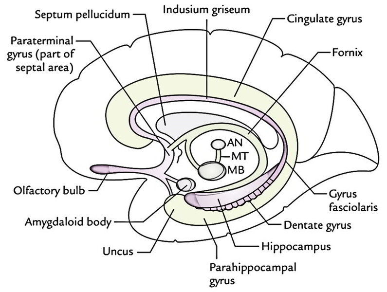
- Limbic lobe, being composed of cingulate gyrus, isthmus, parahippocampal health clubs and uncus (anterior part of the parahippocampal gyrus).
- Hippocampal formation, which consists of hippocampus (cornu ammonis), dentate gyrus, gyrus fasciolaris and indusium griseum.
Subcortical Nuclei
- Amygdaloid nuclear complex (also named amygdaloid body).
- Septal region and nuclei.
- Olfactory areas.
- Hypothalamus especially the mammillary bodies.
- Anterior nucleus of thalamus.
Fibre Bundles of the Limbic System
- Fornix.
- Mammillothalamic tract.
- Stria terminalis.
- Anterior commissure.
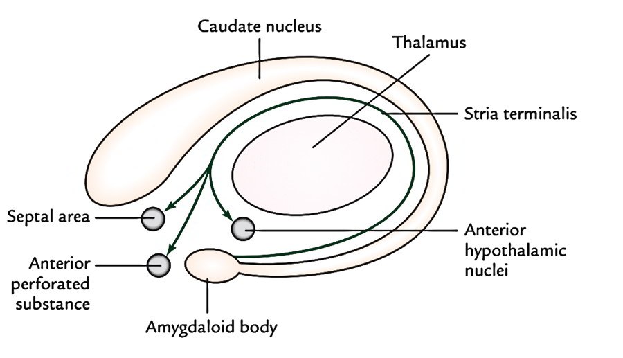
Amygdaloid Nuclear Complex
It is an almond shaped mass of grey matter underlying the rostral part of the parahippocampal gyrus on the anteriormost part of the roof of the inferior horn of lateral ventricle. Amygdaloid nuclear complex includes lateral, central and basal nuclei. Posteriorly, amygdaloid body becomes continuous with the tail of the caudate nucleus and stria terminalis.
Connections
Afferents: Primary afferents to amygdaloid body are from primary olfactory regions. Efferents: Stria terminalis creates the primary efferent tract of the amygdaloid body. It takes a circuitous route together with (but not functionally related to) the tail of caudate nucleus in close relationship to the lateral ventricle until the level of anterior commissure, where bulk of its fibres terminate in the septal area and anterior portion of the hypothalamus. The others join the anterior commissure and are dispersed to the contralateral amygdaloid body. Generally, amygdaloid body plays a significant function in restraining the somatic results to internal demands, drives or instincts. Since part of it gets olfactory stimulation, it’s thought that the amygdaloid body plays a significant role in smellmediated sexual conduct. Arousal of amygdaloid body generates excitability, anxiety and fury. Bilateral damage of amygdaloid body reduces anxiety and increases sexual activity. Folks in late sixties become pervasive in their own sexual behaviour, likely because of atrophy of amygdaloid bodies.
Hippocampal Formation
The hippocampal formation is composed of (a) hippocampus, (b) dentate gyrus, (c) indusium griseum, gyrus fasciolaris and (d) medial and lateral longitudinal striae.
Hippocampus
Hippocampus (also referred to as Ram’s horn or Ammon’s horn) is an area of cerebral cortex that has rolled into the floor of the inferior horn of the lateral ventricle during fetal life. In an adult brain, it creates a longitudinal elevation in the floor of inferior horn of the lateral ventricle and is continuous medially with the subiculum and parahippocampal gyrus. The name “hippocampus” meaning “sea horse”, is originated from its look in coronal section. In the frontal section the hippocampus is C-shaped and its outline bears a resemblance to a Ram’s horn, therefore the name Ram’s horn. It’s also named Ammon’s horn after an Egyptian deity with Ram’s head. Its anterior extremity is enlarged and bears few grooves and intervening ridges. Due to its similarity to a critter’s paw it is called pes hippocampi (pes = foot). Followed posteriorly the hippocampus slowly narrows and finally finishes underneath the splenium of corpus callosum. The ventricular outermost layer of the hippocampus is covered by a thin layer of white fibres referred to as alveus. The fibres of alveus start in the hippocampal cortex, course in the direction of the medial border of hippocampus in the place where they converge to create a narrow strip of white matter, the fimbria of hippocampus. Phylogenetically, hippocampus represents the archicortex and is composed of the following 3 layers:
- Superficial molecular layer.
- Middle pyramidal cell layer.
- Deep polymorphic cell layer.
The parahippocampal cortex (neocortex) is composed of 6 layers. In the region referred to as subiculum, there’s slow transition from 6 -layered neocortex to the 3-layered archicortex.
Connections
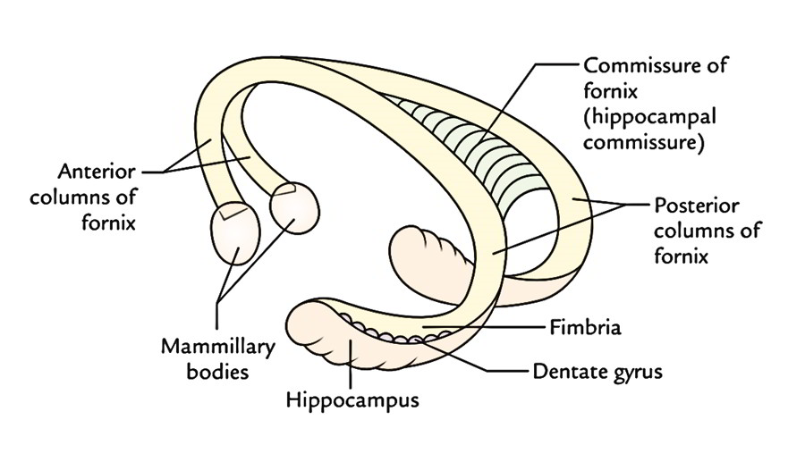
Afferents: Hippocampus gets fibres primarily from entorhinal area (area 28). Efferents: The fornix is the primary efferent tract of the hippocampus. The fibres making the hippocampus pass to:
- The opposite hippocampus via the commissure of fornix/hippocampal commissure,
- The septal and anterior hypothalamic regions and
- The mammillary body, which sends nerve impulses to cingulate gyrus via anterior nucleus of thalamus.
Functions of Hippocampus
Once hippocampus was regarded as the part of olfactory system but it’s no direct connections with the sense of smell in man. In man it’s an integrative center, which affects endocrine and visceral functions and emotional states via its links with hypothalamus, septal nuclei and the cingulate gyrus. It plays an essential job in recent memory.
Dentate Gyrus, Indusium Griseum and Medial and Lateral Longitudinal Striae
In the fetal brain, the dentate gyrus grows as a further expansion of the hippocampus and takes up the period between the hippocampus and the parahippocampal gyri, being located deep to fimbria. Its surface is toothed, for this reason the name dentate gyrus. When followed anteriorly, dentate gyrus runs medially across the inferior surface of the uncus. This part is known as tail of dentate gyrus. The posterior end of dentate gyrus is constant with the splenial gyrus or gyrus fasciolaris, which continues as a thin layer of grey matter over the corpus callosum named indusium griseum. The indusium griseum is the vestigial grey matter and includes 2 fine longitudinal bands of fibres buried in it, the medial and lateral longitudinal striae.
Fornix
The fornix is a large bundle of projection fibres, which attaches the hippocampus with the mammillary body. It makes up the only efferent system of the hippocampus.
 On the medial surface of the cerebral hemisphere, it’s viewed as an arched notable bundle of white fibres below the corpus callosum, along the lower border of septum pellucidum. There’s 1 fornix in every cerebral hemisphere but 2 are thus closely related/fused below the middle of the body of corpus callosum they are generally defined as a single structure.
Origin, Course and Distribution of the Fibres
The fibres of fornix originate primarily from the pyramidal cells of the hippocampus and create a thin layer of white fibres on its ventricular surface referred to as alveus. The fibres of alveus pile on the medial margin of hippocampus to create a narrow strip of white matter, the fimbria, being located flat over the dentate gyrus. The fimbria becomes a rounded band, the crus of fornix as it arches upwards, medially and forwards underneath the splenium of corpus callosum. Both crura, 1 of every hemisphere, arch over the thalamus, converge and connect in the midline underneath the trunk of corpus callosum to create the body of fornix. Anteriorly, the body of fornix breaks up into 2 columns, the columns of fornix. Every column of fornix arches downwards in the direction of the anterior commissure and creates the anterior boundary of interventricular foramen. Subsequently it arches posteriorly via the hypothalamus to finish in the mammillary body. These fibres being located posterior to anterior commissure are called postcommissural fornix. Some fibres of column pass in front of anterior commissure to finish in the septal area and anterior hypothalamic region, etc. to make up the precommissural fornix. To summarize, the principal parts of the fornix are fimbria, crura, body and anterior columns. Fornix is the only tract of the cerebrum, which includes all the 3 types of its fibres like projection fibres, commissural fibres and association fibres.
Mammillothalamic Tract
Mammillothalamic tract is a notable bundle of fibres, also termed, Felix Vicq d’Azyr, which carry nerve impulses from mammillary body to the anterior, nucleus of the thalamus.

 (52 votes, average: 4.63 out of 5)