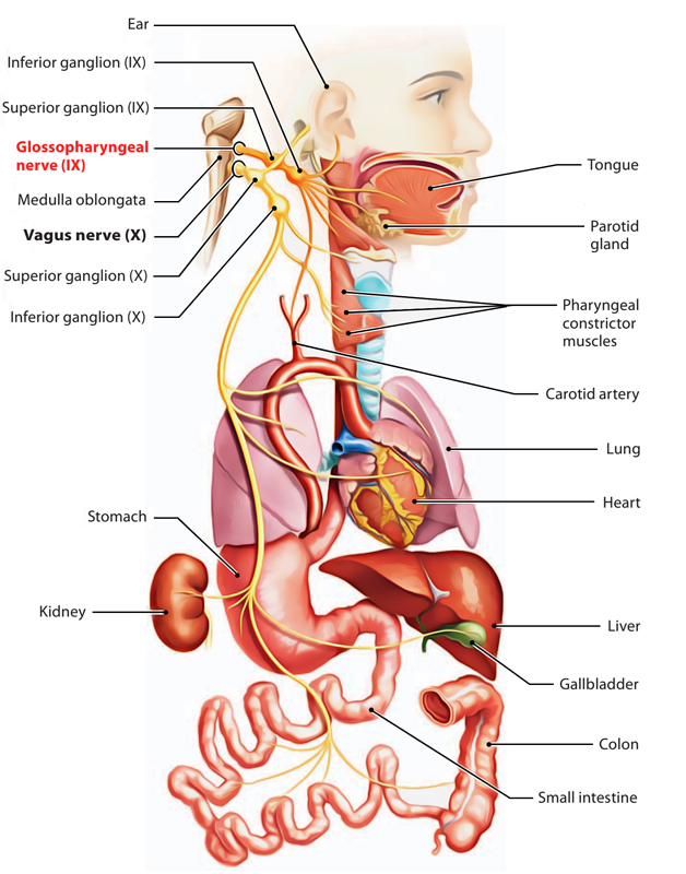
It’s a mixed nerve, i.e., composed of both the motor and sensory fibres but mainly it’s sensory. Glossopharyngeal nerve is the 9th cranial nerve. It derives its name from the truth that it gives sensory innervation to the tongue and pharynx.

Glossopharyngeal Nerve
Functional Parts and Nuclei
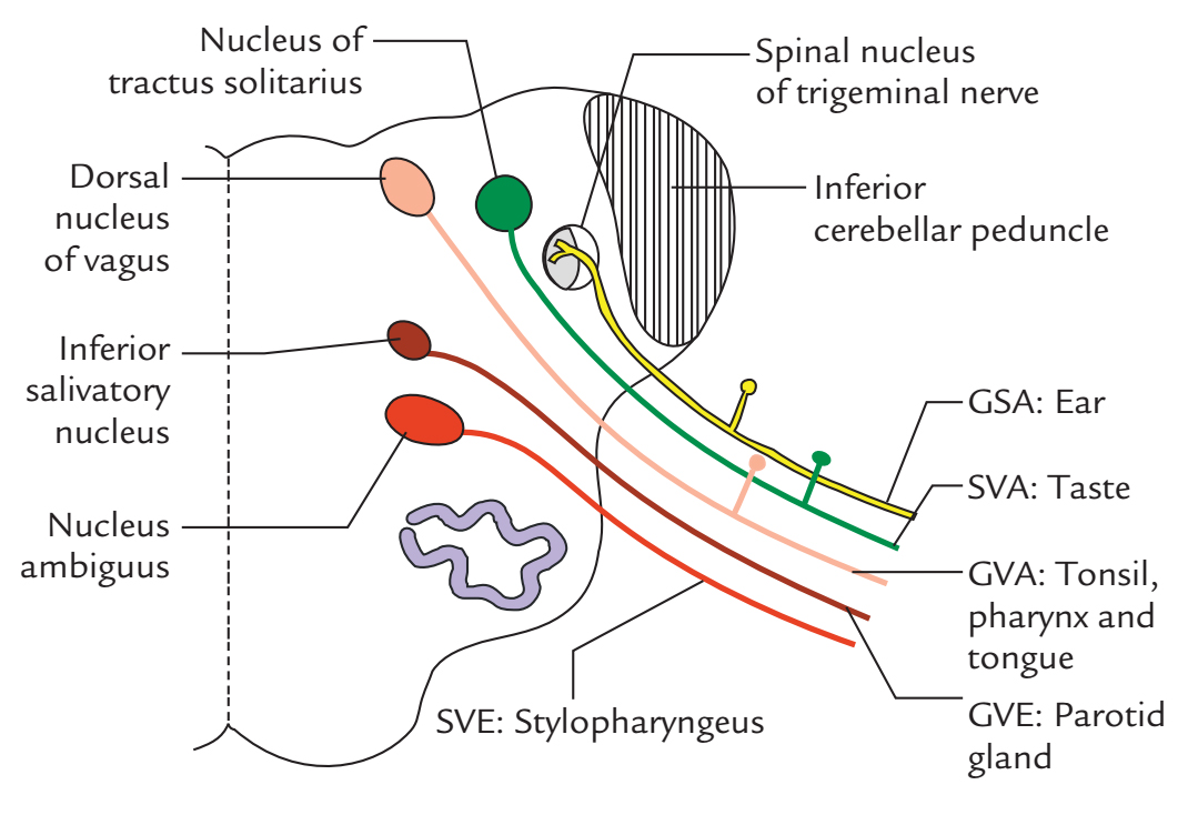
- Special visceral efferent fibres: They supply the stylopharyngeus muscle. They originate from nucleus ambiguus.
- Special visceral afferent fibres: They carry taste sensations from the posterior one-third of tongue consisting of vallate papillae and terminate in the nucleus tractus solitarius.
- General visceral efferent fibres: They supply the secretomotor fibres to the parotid gland They originate from the inferior salivatory nucleus and are preganglionic parasympathetic fibres.
- General visceral afferent fibres: They carry general sensations of pain, feel and temperature from the mucous membrane of the pharynx, tonsil, soft palate and the posterior one-third of tongue and terminate in the dorsal nucleus of the vagus.
- General somatic afferent fibres: They carryproprioceptive sensations from the stylopharyngeus and skin of the auricle and terminate in the nucleus of the spinal tract of 5th nerve.

Glossopharyngeal Nerve: Functional Parts and Nuclei
Course and Connections
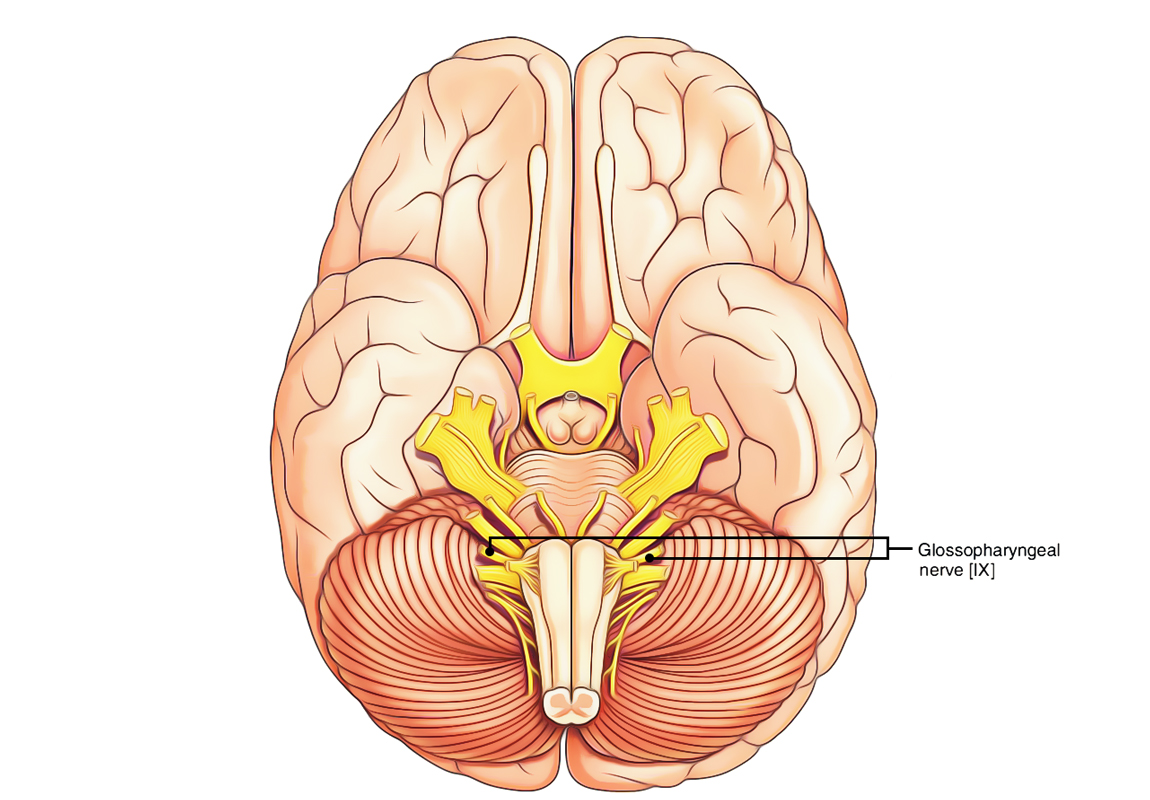
- The glossopharyngeal nerve originates from the upper part of the lateral aspect of the medulla between the olive and the inferior cerebellar peduncle by three or four rootlets. The rootlets unify to create just one trunk which runs forwards and laterally to leave the cranial cavity by going through the intermediate compartment of the jugular foramen enclosed in another sheath of dura mater.
Glossopharyngeal Nerve: Course and Connections
- The superior and inferior sensory ganglia are on the nerve as it goes through the jugular foramen.
- The smaller superior ganglion is located inside the jugular foramen and is regarded as the separated part of the inferior ganglion.
- The bigger inferior ganglion is located just below the jugular foramen and includes the cell bodies of majority of the sensory fibres of the nerve.
- After appearing from the jugular foramen at the base of the skull, the nerve enters downward and forwards between the internal carotid artery and the internal jugular vein. It then descends anterior to the internal carotid artery to the styloid process and muscles connected to it (in this position, it is located lateral to the tonsillar bed created by the superior constrictor) to reach the lower border of the stylopharyngeus. From here it enters together with the stylopharyngeus via the gap between the superior and middle constrictors of the pharynx.
- It then arch forwards along the lateral aspect of the stylopharyngeus muscle which it supplies and after that enters deep to the stylohyoid ligament and posterior border of the hyoglossus muscle. Here it breaks up into terminal branches, which supply the mucous membrane of the posterior 1 third of the tongue, pharynx and tonsil.
Branches and Distribution
The glossopharyngeal Nerve Supplies the following essential branches:
Tympanic branch (Jacobson’s nerve)
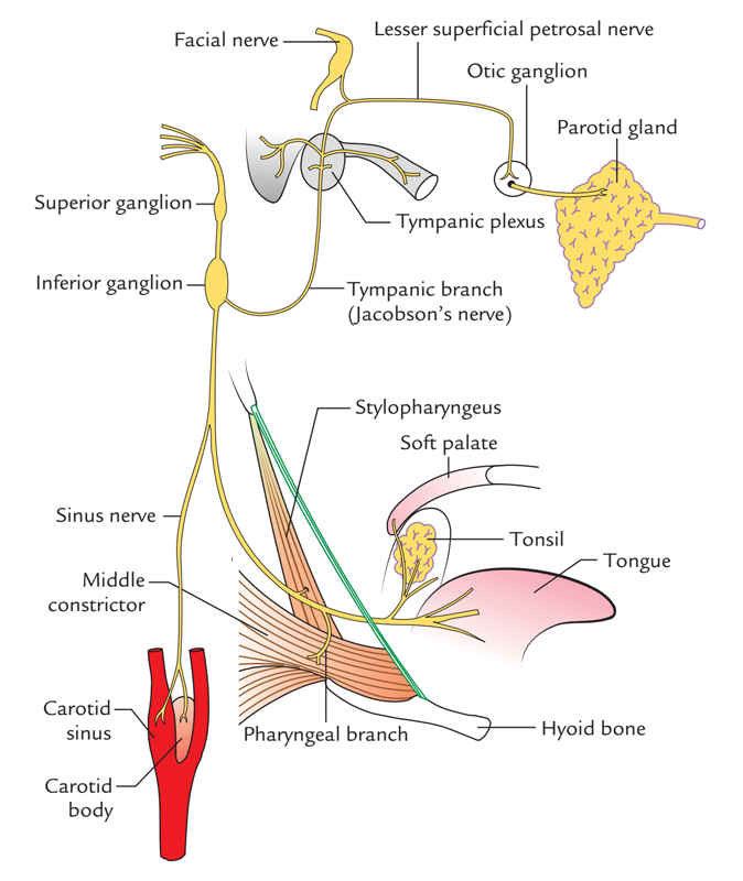
It leaves the inferior ganglion and enters the middle ear via the tympanic canaliculus situated in the bony border between the jugular foramen and carotid canal. It creates the tympanic plexus over the promontory of the middle ear.
The tympanic plexus produces the lesser petrosal nerve and twigs to tympanic cavity, auditory tube and mastoid air cells.

Glossopharyngeal Nerve: Branches and Distribution
The lesser petrosal nerve carries the preganglionic parasympathetic fibres which relay in the otic ganglion. The postganglionic fibres from the ganglion supply the parotid gland
Carotid nerve (nerve of Herring)
It’s a branch to carotid sinus and carotid body. It acts as an afferent limb for pressoreceptor and chemoreceptor reflexes from the carotid sinus and carotid body to regulate the pulse and respiration, respectively.
Pharyngeal branch
It joins the pharyngeal branches of the vagus and the cervical sympathetic chain to create the pharyngeal plexus on the middle constrictor of the pharynx
Branch to stylopharyngeus
It appears as the nerve, winds round the stylopharyngeus muscle. It’s the only motor branch of the glossopharyngeal nerve.
- Tonsillar branches: They supply the mucous membrane of tonsil, fauces and palate.
- Lingual branches: They supply the posterior one-third of the tongue and vallate papillae and express flavor and general sensations.
Clinical Significance
Lesions of Glossopharyngeal Nerve
The lesion of the glossopharyngeal nerve is uncommon in isolation since there’s frequently related engagement of the vagus nerve. Yet, the complete lesion of the glossopharyngeal nerve results in:
- The loss of flavor and general sensations over the posterior one-third of the tongue,
- Trouble in swallowing,
- The decrease of the salivation from the parotid gland and
- The unilateral reduction of the gag reflex.
Clinical Testing of Glossopharyngeal Nerve
Theglossopharyngeal nerve can be examined medically by:
- Evoking the gag reflex (i.e., on tickling the posterior wall of the pharynx, soft palate, or tonsillar fossa, there’s reflex contraction of pharyngeal muscles causing gagging and retching) and
- Examining the taste sensations in the posterior one-third of the tongue.
Glossopharyngeal Neuralgia
Glossopharyngeal neuralgia, in spite of the fact that uncommon, may happen. It’s characterized by paroxysmal episodes of intractable pain in the area of the sensory distribution of the glossopharyngeal nerve, example, throat, tongue and ear, precipitated by consuming.


 (56 votes, average: 4.59 out of 5)