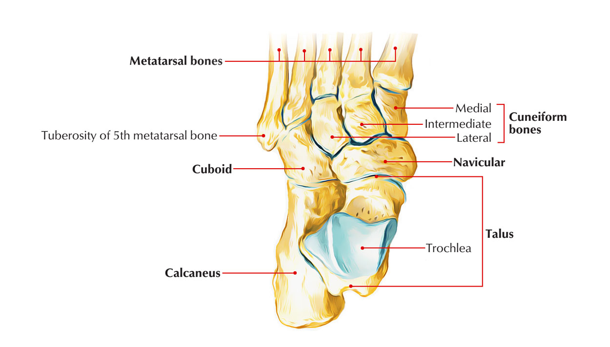
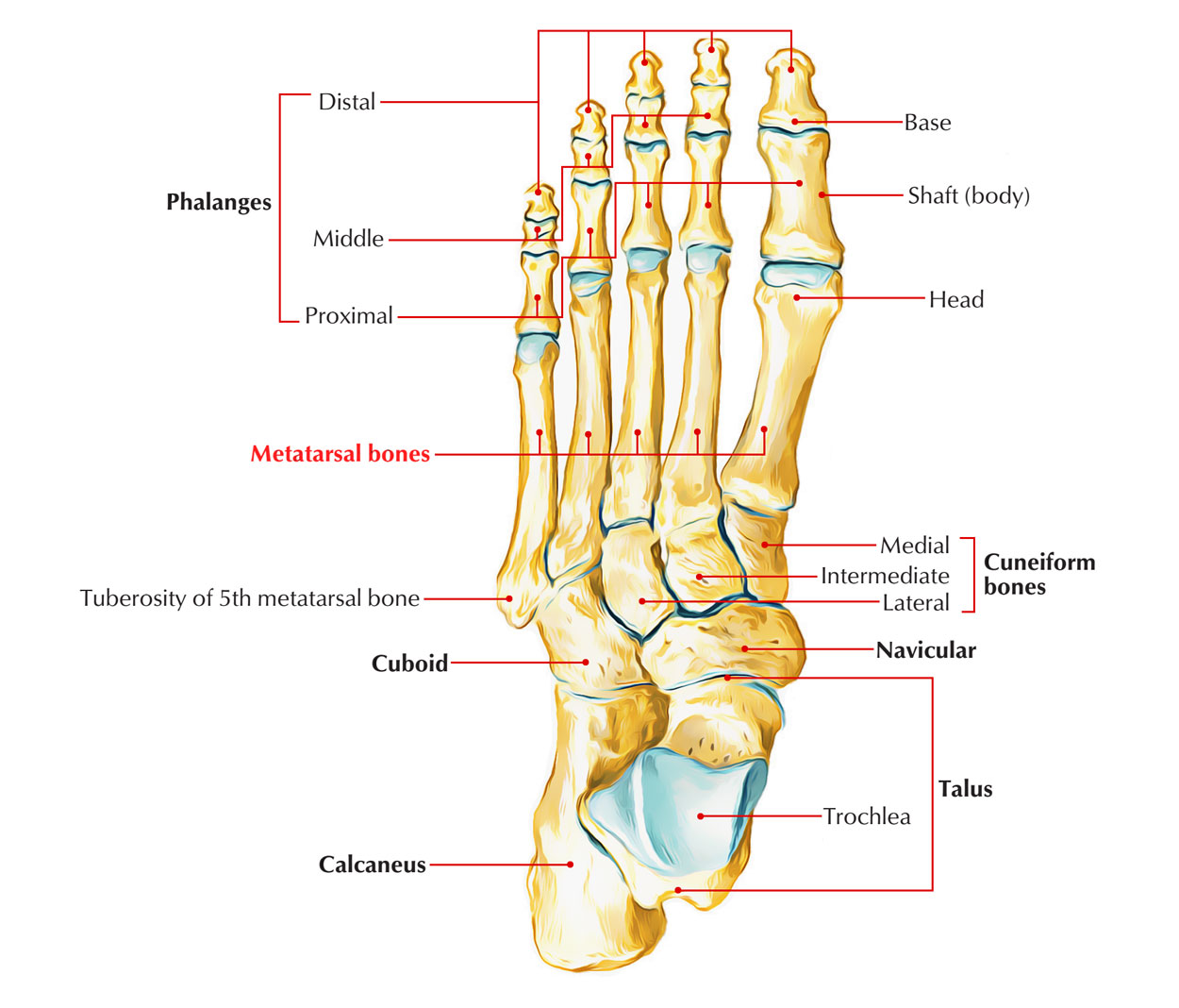
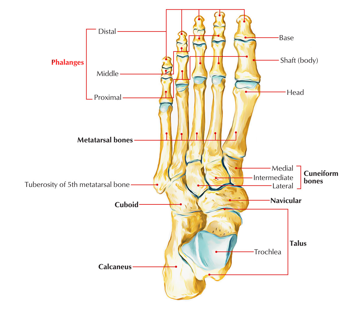
The skeleton of the foot from behind forward is composed of the following bones:
- Tarsals.
- Metatarsals.
- Phalanges.
Tarsal Bones
All these are short bones which collectively create tarsus. These arearranged in 3 rows:
(a) Proximal row includes talus and calcaneus.
(b) Middle row is made of navicular.
(c) Distal row is composed of 3 cuneiforms (medial, intermediate, and lateral) and cuboid.

Skeleton of the Foot: Tarsal Bones
Identification of bones in the skeleton of the foot:
- Calcaneus (heel bone) is the largest and most proximal bone.
- Talus is the 2nd largest bone and is located above the calcaneus like a rider, therefore maximum bone in the skeleton of foot.
- Navicular is boat-shaped and is located in front of the head of talus.
- Cuboid is cubical in shape in front of the lateral part of calcaneum.
- Cuneiforms are small wedge shaped bones and ordered from side to side in front of navicular.
Metatarsal Bones
All these are 5 tiny long bones. The 5 metatarsal bones collectively make up the metatarsus. They can be numbered from medial to lateral sides as first, second, third, fourth, and fifth.

Skeleton of the Foot: Metatarsal Bones
Identification
First Metatarsal
- It’s the shortest, thickest, and most powerful, and is accommodated for weight transmission.
- Proximal surface of its base presents a kidney shaped articular surface.
Second Metatarsal
- It’s the longest metatarsal bone.
- Proximal surface of its base has a triangular concave articular surface.
Third Metatarsal
- Proximal surface of its base has a flat triangular articular facet.
- The lateral side of its base has 2 facets while the medial side has 1 facet.
Fourth Metatarsal
- The proximal surface of its base has a quadrilateral facet, which articulates with the cuboid.
- The lateral side of base has 1 facet while its medial side has 1 facet split into 2 parts- proximal and distal.
Fifth Metatarsal
The lateral side of its base projects proximally and somewhat laterally to create a large tuberosity (styloid process).
Phalangeal Bones
The phalangeal bones are tiny long bones. They’re 14 in number in every foot- 2 for the great toe and 3 for every of the other 4 toes. The phalanges in the great toe are proximal and distal, and phalanges in other toes are proximal, middle, and distal.

Skeleton of the Foot: Phalangeal Bones
- The base of the proximal phalanx presents a concave facet which joint together with the head of metatarsal bone to create the metacarpophalangeal joint.
- The distal end of the proximal phalanx presents a pulley like articular surface and articulates with the middlephalanx in the lateral 4 toes and with terminal phalanx of first toe. So, lateral 4 toes possess 2 interphalangeal (IP) joints, proximal and distal, and the great toe possesses only 1 IP joint.
- Both, proximal and distal articular surfaces of the middle phalanx are pulley-shaped.
- The distal phalanx of every toe bears a rough tuberosity on plantar aspect of its distal end.
Ossification
- Every phalanx ossifies from 2 centers: a primary center for the shaft and a secondary center for the base.
- The period of appearance of primary centers is as follows:
(a) For proximal phalanges: 12th week of IUL (Intrauterine Life).
(b) For middle phalanges: 15th week of IUL.
(c) For distal phalanges: 9th week of IUL. - The period of appearance of secondary centers is as follows:
(a) For proximal phalanges: 2 years.
(b) For middle phalanges: 4 years.
(c) For distal phalanges: 8 years. - The fusion of epiphysis (base) with diaphysis (shaft) takes place in about the 18th year.

 (54 votes, average: 4.87 out of 5)