The heart is provided by the sympathetic and parasympathetic fibres via the superficial and deep cardiac plexuses created by the parasympathetic and sympathetic fibres.
The parasympathetic fibres are originated from vagus nerves. They’re cardioinhibitory, therefore, their stimulation causes slowing of the heart rate and constriction of the coronary arteries.
The sympathetic fibres are originated from upper 35 thoracic spinal segments. They’re cardioacceleratory, for this reason their stimulation increase the heart rate and causes the dilatation of the coronary arteries. The sympathetic fibres also cause dilatation of the coronary arteries.
A brief account of formation and distribution of cardiac plexuses is provided in the following text.
Cardiac Plexuses
Superficial Cardiac Plexus
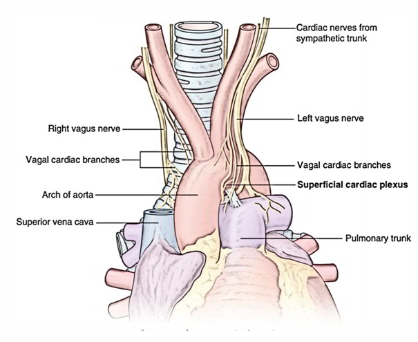
The superficial cardiac plexus is located below the arch of aorta in front of the bifurcation of pulmonary trunk, just to the right of ligamentumarteriosum. The cardiac ganglion (of Wrisberg) is located close to the ligamentumarteriosum.
It’s created by the:
A. Superior cervical cardiac branch of left cervical sympathetic trunk, and
B. Inferior cervical cardiac branch of left vagus nerve.
Distribution
The superficial cardiac plexus gives branches to (a) deep cardiac plexus, (b) right coronary artery, and (c) left anterior pulmonary plexus.
Deep Cardiac Plexus
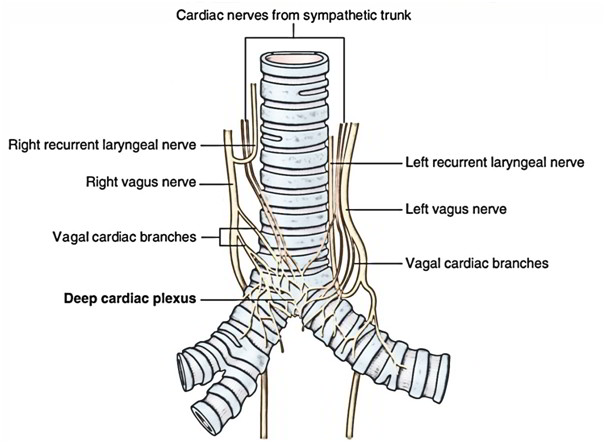
The deep cardiac plexus is located in front of the bifurcation of the trachea, behind the arch of the aorta.
It’s created by:
A. all the cardiac branches originated from 3 cervical and upper 4 or 5 thoracic ganglia of the sympathetic chains with the exception of the superior cervical cardiac branch of left cervical sympathetic chain, and.
B. all the cardiac branches of vagus and recurrent laryngeal nerves with the exception of the inferior cervical cardiac branch of the left vagus nerve.
Distribution
The left and right halves of the plexus distributes branches to (a) corresponding coronary arteries and pulmonary plexus, and (b) separate branches to the atria.

 (56 votes, average: 4.55 out of 5)