Quadriceps Femoris is a muscle and is thus referred to as because it is composed of 4 parts: rectus femoris, vastus lateralis, vastus medialis, and vastus intermedius. It’s supplied by the femoral nerve. It creates the majority of the mass on the anterior aspect of the thigh and is the strongest extensor of the knee joint.
All the parts of quadriceps cross only 1 joint, i.e., knee joint, with the exception of rectus femoris which crosses 2 joints, i.e., hip and knee joints.
The origin, insertion, and Nerve Supply of different parts of quadriceps femoris are explained in the subsequent text.
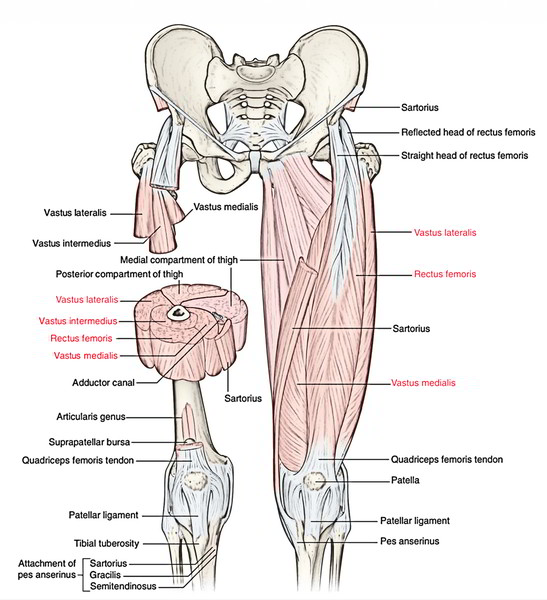
Rectus Femoris
It’s a long straight muscle with fusiform bipinnate abdomen in the upper two-third and flat tendon in the lower one-third (Latin rectus = straight).
Origin
It originates from the ilium by 2 heads:
- Straight head originates from the upper part of the anterior inferior iliac spine.
- Reflected head appears from a groove above the acetabulum. Insertion.
- The 2 heads meet at an acute angle and create a fusiform abdomen, which takes up the front of the thigh. In the lower one-third of the thigh, the muscle ends in a flat tendon which descends vertically downward to be added into the base of the patella.
Nerve Supply
It’s supplied by the posterior section of the femoral nerve.
Actions
It stretches the knee and bends the hip.
The rectus femoris is also termed the “kicking muscle” because it stretches the knee and bends the hip- the Actions needed during kick.
Vastus Lateralis
Origin
It gets a linear and aponeurotic origin from the upper part of the intertrochanteric line, anterior and inferior edges of the higher trochanter, lateral lip of gluteal tuberosity, and upper half of the lateral lip of linea aspera. It also originates from the lateral intermuscular septum.
Insertion
It’s added (a) by a broad aponeurosis into the tendon of rectus femoris, lateral part of the base of patella, upper one-third of the lateral border of the patella, and (b) retinacular fibres to the very front of the lateral condyle of tibia which replaces the anterolateral aspect of the capsule of the knee joint.
Nerve Supply
It’s supplied by the posterior section of the femoral nerve.
Actions
Extension of the knee joint.
Vastus Medialis
It covers the medial surface of the shaft of femur.
Origin
It requires a linear origin from the lower part of intertrochanteric line, coil line, medial lip of linea aspera, and upper two-third of the medial supra condylar line. It also originates from the medial intermuscular septum.
Insertion
It’s fit by a broad aponeurosis into the tendon of rectus femoris, medial part of the base, and upper two-third of the medial border of the patella. From its lower connection it sends a fibrous growth, the medial patellar retinaculum, to the very front of the medial condyle of the tibia which replaces the anteromedial part of the capsule of the knee joint.
Nerve Supply
It’s supplied by the nerve to vastus medialis, that is the thickest muscular branch of the posterior section of the femoral nerve.
Actions
Expansion of the knee joint.
The lower fibres of vastus medialis are connected much more down on the medial border of patella than those of the vastus lateralis on the lateral border. Consequently, they prevent the natural inclination of patella to dislocate laterally during the extension of the knee joint.
The function of vastus medialis is essential for the stability of the patella.
Vastus Intermedius
Origin
It originates from the anterior and lateral aspects of the upper three-fourth of the shaft of femur.
Insertion
Into the base of patella deep to the tendon of rectus femoris.
Nerve Supply
It’s supplied by the posterior section of the femoral nerve.
Actions
Extension of the knee joint.

 (58 votes, average: 4.88 out of 5)