The ear is the organ of hearing. It is also the organ of equilibrium. The ear is subdivided into three major parts: the external ear, middle ear, and internal ear.

External Ear
The external ear consists of two parts: the auricle and the external acoustic meatus. The auricle (pinna) is the funnellike structure composed primarily of cartilage and skin that is attached to the side of the head. The external acoustic meatus is a short tube that extends from the auricle through the temporal bone to the eardrum. Sound waves striking the auricle are channeled into the external acoustic meatus. Cerumen (earwax) and hairs in the external acoustic meatus help to prevent foreign particles from reaching the eardrum.
Middle Ear
The middle ear, or tympanic (tim-pan’-ik) cavity, is an air-filled space within the temporal bone. The tympanic membrane, auditory tube, and auditory ossicles are parts of the middle ear. The tympanic membrane, or eardrum, separates the tympanic cavity from the external acoustic meatus. The tympanic membrane is covered with skin externally and by a mucous membrane internally. Sound waves, or air pressure waves, entering the external acoustic meatus cause the tympanic membrane to vibrate in and out at the same frequency as the sound waves.
The auditory (eustachian) tube connects the tympanic cavity with the pharynx. Its function is to keep the air pressure within the tympanic cavity the same as the external air pressure by allowing air to enter or exit the tympanic cavity. Equal air pressure on each side of the tympanic membrane is essential for the tympanic membrane to function properly. A valve at the pharyngeal end of the tube is usually closed but it opens when a person swallows or yawns to allow air pressure to equalize. If you have experienced a rapid change in air pressure, you probably have noticed your ears “popping” as the air pressure is equalized and the tympanic membrane snaps back into place.
The auditory ossicles (os’-si-kulz) are three tiny bones that articulate to form a lever system from the tympanic membrane, across the tympanic cavity, to the internal ear. Each ossicle is named for its shape. The tip of the “handle” of the club-shaped malleus (mal’-e-us), or hammer, is attached to the tympanic membrane and its head articulates with the incus (ing’-kus), or anvil. The base of the incus articulates with the stapes (sta’-pez), or stirrup, whose foot plate is inserted into the oval window of the internal ear.
The vibrations of the tympanic membrane cause corresponding movements of the ossicles, which result in the stapes vibrating in the oval window. In this way, vibrations of the tympanic membrane are transmitted to the fluid-filled internal ear. Due to the size difference between the larger tympanic membrane and the smaller oval window, vibrations are amplified by the ossicles.
Internal Ear
The internal ear is embedded in the temporal bone. It consists of two series of connecting tubes and chambers, one within the other: an external bony labyrinth (lab’- i-rinth) and an internal membranous labyrinth. The two labyrinths are similar in shape. The space between the bony and membranous labyrinths is filled with perilymph, whereas the membranous labyrinth contains endolymph. These fluids play important roles in the functions of the internal ear. The internal ear has three major parts: the cochlea, vestibule, and semicircular canals.
The cochlea (kok’-le-ah) is the coiled portion of the internal ear. When viewed in cross section, the cochlea is composed of three chambers that are separated from each other by membranes. The scala vestibuli (ska-la ves-tib ‘-u-li) and the scala tym- pani, both components of the bony labyrinth, extend the length of the cochlea and are continuous with each other at the apex of the cochlea. The scala vestibuli continues into the vestibule, which houses the membrane-covered oval window. The scala tympani extends toward the vestibule, ending at the membrane-covered round window.
The cochlear duct, which is part of the membranous labyrinth, extends nearly to the apex of the cochlea. It is separated from the scala vestibuli by the vestibular membrane and from the scala tympani by the basilar membrane. The basilar membrane contains about 20,000 cross fibers that gradually increase in length from the base to the apex of the cochlea. The attachment of the basilar membrane to the bony center of the cochlea allows it to vibrate like the reeds of a harmonica when activated by vibrations generated by sound.
The spiral organ (organ of Corti), which contains the sensory receptors for sound stimuli, supported by the basilar membrane within the cochlear duct. The sensory receptors are called cochlear hair cells, and they have hairlike stereocilia extending from their free surfaces toward the overlying tectorial (tek-to’-re-al) membrane. Axons of the cochlear branch of the vestibulocochlear nerve (CN VIII) exit the cochlear hair cells and lead to the brain.
Ear Functions
| Structure | Function |
| External Ear | |
| Auricle | Channels sound waves into external acoustic meatus |
| External acoustic meatus | Directs sound waves to tympanic membrane |
| Tympanic membrane Middle Ear | Vibrates when struck by sound waves |
| Tympanic cavity | Air-filled space that allows tympanic membrane to vibrate freely when struck by sound waves |
| Auditory ossicles | Transmit and amplify vibrations produced by sound waves from the tympanic membrane to the perilymph within the cochlea |
| Auditory tube Internal Ear | Equalizes air pressure on each side of tympanic membrane |
| Cochlea | Fluids and membranes transmit vibrations initiated by sound waves to the spiral organ, whose cochlear hair cells generate nerve impulses associated with hearing |
| Saccule | Vestibular hair cells of the macula form nerve impulses associated with static and dynamic equilibrium |
| Utricle | Vestibular hair cells of the macula form nerve impulses associated with static and dynamic equilibrium |
| Semicircular canals | Vestibular hair cells of the crista ampullaris form nerve impulses associated with dynamic equilibrium |
Physiology of Hearing
The human ear is able to detect sound waves with frequencies ranging from near 20 to 20,000 Hertz (Hz; vibrations per second), but hearing is most acute between 2,000 and 3,000 Hz. For hearing to occur, vibrations formed by sound waves must be transmitted to the cochlear hair cells of the spiral organ. Then, the cochlear hair cells form nerve impulses that are transmitted to the hearing areas of the cerebrum for interpretation as sound sensations.
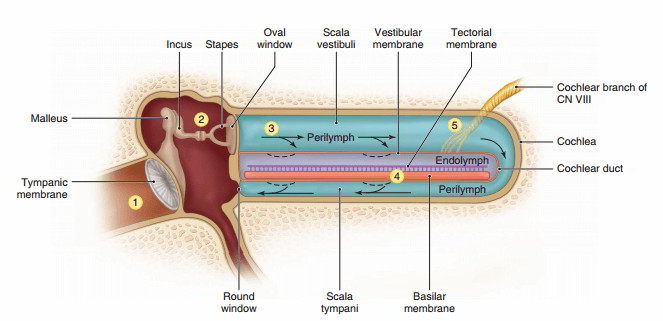
Figure above shows the structure of the internal ear with the cochlea uncoiled to show more clearly the relationships of its parts. Refer to this figure as you study the following outline of hearing physiology.
- Sound waves enter the external acoustic meatus and strike the tympanic membrane, causing it to vibrate in and out at the same frequency and comparable intensity to the sound waves. Loud sounds cause a greater displacement of the tympanic membrane than do soft sounds.
- Vibration of the tympanic membrane causes movement of the auditory ossicles, resulting in the in-and-out vibration of the stapes in the oval window.
- The vibration of the stapes causes a corresponding oscillatory (back-and-forth) movement of the perilymph in the scala vestibuli and scala tympani and a corresponding movement of the membrane over the round window. This movement of the perilymph causes vibrations in the vestibular and basilar membranes.
- The vibration of the basilar membrane causes the stereocilia of the cochlear hair cells to contact the tectorial membrane, which stimulates the formation of nerve impulses by the cochlear hair cells.
- Nerve impulses formed by the cochlear hair cells are carried by the cochlear branch of the vestibulocochlear nerve to the hearing areas of the temporal lobes of the cerebrum, where the sensation is interpreted. Some of the axons cross over to the opposite side of the brain so that the hearing areas in each temporal lobe interprets nerve impulses originating in each ear. APlR
Pitch And Loudness
Because of the gradually increasing length of the fibers in the basilar membrane, different portions of the basilar membrane vibrate in accordance with the different frequencies (pitch) of sound waves. Low-pitched sounds cause the longer fibers of the membrane near the apex of the cochlea to vibrate, and high-pitched sounds activate the shorter fibers of the membrane near the base of the cochlea. The pitch of a sound sensation is determined by the portion of the basilar membrane and the spiral organ that are activated by the specific sound frequency and by the parts of the hearing areas that receive the nerve impulses. Nerve impulses from different regions of the spiral organ go to slightly different portions of the hearing areas in the brain, which causes them to be interpreted as different pitches.
The loudness of the sound is dependent upon the intensity of the vibration of the basilar membrane and spiral organ, which, in turn, determines the frequency of nerve impulse formation. The greater the frequency of nerve impulses sent to the brain, the louder the sound sensation.
Disorders of The Ear
Deafness is a partial or total loss of hearing. The cochlear hair cells of the spiral organ are easily damaged by high- intensity sounds, such as loud music and the noise of jet airplanes. Such damage produces a form of nerve deafness that may be partial or total, and it is permanent. Disorders of sound transmission by the tympanic membrane or auditory ossicles cause conduction deafness, which may be repairable by surgical means or overcome by the use of hearing aids.
Labyrinthine disease is a term applied to disorders of the internal ear that produce symptoms of dizziness, nausea, ringing in the ears (tinnitis), and hearing loss. It may be caused by an excess of endolymph, infection, allergy, trauma, circulation disorders, or aging.
Motion sickness is a functional disorder that is characterized by nausea and is produced by repetitive stimulation of the equilibrium receptors in the internal ear.
Otitis media (o-ti ‘-tis me ‘-de-ah) is an acute infection of the tympanic cavity. It may cause severe pain and an outward bulging of the tympanic membrane due to accumulated fluids. Pathogens enter the middle ear from the pharynx via the auditory tube or through a perforated tympanic membrane. Young children are especially susceptible because their auditory tubes are short and horizontal, which aids the spread of bacteria from the pharynx to the tympanic cavity.

 (60 votes, average: 4.68 out of 5)