Lymphoid structures can be found throughout the body. While all lymphoid structures are capable of lymphocyte production, the red bone marrow and thymus are considered primary lymphoid organs because all WBCs, especially lymphocytes, originate in these organs. After production in the red bone marrow most lymphocytes and other immune cells go to secondary lymphoid organs, such as the lymph nodes and spleen that become the sites of proliferation of lymphocytes and immune responses.
Red Bone Marrow
Red bone marrow is hematopoietic (blood-forming) tissue found in the spongy bone of most of the axial skeleton and the proximal epiphyses of the humerus and femur. Red bone marrow is the site of origin of all formed elements in the blood. Not all lymphocytes formed in the red bone marrow are immunocompetent, capable of recognizing and attacking foreign antigens, when they exit the marrow. To be immunocompetent, a lymphocyte must be able to elicit an immune response. B cells, or B lymphocytes, stay in the bone marrow until they are immunocompetent before moving on to secondary lymphoid organs. Other lymphocytes, however, must first move to the thymus for maturation before moving to secondary lymphoid organs.
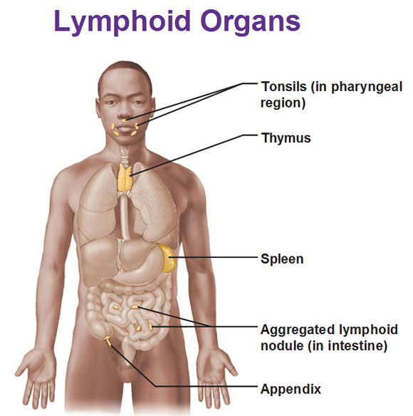
Thymus
The thymus is a soft, bilobed gland located in the mediastinum superior to the heart. It is large (40 g) in infants and children, but after puberty it begins to atrophy and becomes quite small (12 g) in adults. The thymus plays a key role in the development of the lymphoid system before birth and during early childhood. Until the lymphoid system matures at about two years of age, an infant is more susceptible to disease than older children. The major function of the thymus is the differentiation of a class of lymphocytes called T cells, or T lymphocytes, into immunocompetent cells. The thymus produces hormones called thymosins that promote the differentiation and division of T cells, making them immunocompetent. After maturation, T cells are distributed by the blood to secondary lymphoid organs and lymphoid tissues throughout the body.
Lymph Nodes
Lymph nodes usually occur in groups along the larger lymphatic vessels. They are widely distributed in the body, but they do occur in large collections in the inguinal, axillary, and cervical regions of the body, as well as within the ventral cavity. There are no lymph nodes in the CNS.
Structure of Lymph Nodes
Lymph nodes are roughly bean-shaped and 1.0 to 2.5 cm in length. Note that the lymph node consists of a number of small subunits called lymphoid nodules. The lymphoid nodules are collections of lymphocytes and macrophages within reticular tissue and are the sites of activation and proliferation of lymphocytes.
Lymph enters a lymph node through several afferent (af-er-ent) lymphatic vessels and flows through the lymphatic sinuses, which surround the lymphoid nodules. Lymph is collected from the sinuses and enters efferent (ef-er-ent) lymphatic vessels, which carry lymph away from the lymph node. The indentation of the node where efferent lymphatic vessels emerge is called the hilum.
Function of Lymph Nodes
A major function of lymph nodes is the filtration and cleansing of the lymph as it passes through a node. They are the only lymphoid organs that filter the lymph. Damaged cells, cancerous cells, cellular debris, bacteria, and viruses become trapped in the reticular tissue of the lymph node and are destroyed by the action of lymphocytes and macrophages. Lymphocytes act against cancerous cells and pathogens, such as bacteria and viruses. Macrophages engulf cellular debris, immobilized or dead bacteria, and viruses.
Spleen
The spleen is the largest lymphoid organ. It is located posterior to the stomach near the diaphragm in the left upper quadrant of the abdominopelvic cavity. The false ribs provide protection against physical injury. The spleen is a soft, purplish organ 5 to 7 cm (2-3 in) wide and 13 to 16 cm (5-6 in) long. It contains numerous centers for lymphocyte proliferation and a large venous sinus filled with blood.
The spleen resembles a large lymph node. Like lymph nodes, it is enveloped by a thin capsule of dense irregular connective tissue and is subdivided by reticular tissue into many compartments. The compartments contain two basic types of tissues that are named for their appearance in fresh, unstained tissue. White pulp consists of large numbers of lymphocytes that cluster around tiny branches of the splenic artery. This tissue is mostly concerned with the immune functions of the spleen. Red pulp occupies the rest of a compartment, surrounding the white pulp and the venous sinuses. It is a storage area for formed elements and a site where worn-out red blood cells and pathogens are removed from the blood. Before birth, the spleen and liver are the major bloodforming organs, but this function is later taken over by red bone marrow. After birth, the spleen’s role is related to both lymphoid and cardiovascular functions:
- It cleanses and filters the blood much like lymph nodes cleanse lymph. Lymphocytes and macrophages destroy pathogens, and macrophages clean up the debris.
- It stores a reserve supply of RBCs and platelets, which can be released into the blood in times of need, such as after a hemorrhage.
- It is a major site for RBC destruction and recycling.
- It is a major site of lymphocyte activation and proliferation.
In spite of these important functions, the spleen is not essential for life. But following a splenectomy, a person may be more susceptible to potential pathogens and the effects of hemorrhage.

 (51 votes, average: 4.67 out of 5)