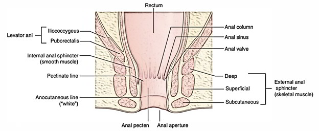
The external anal sphincter, which covers the anal canal, is composed of three portions deep, superficial, along with subcutaneous throughout the canal from superior to inferior organized sequentially and is created by skeletal muscle.

Anal Sphincter
Parts
Deep Part
The deep part circles around the upper portion of the anal canal and mixes along with the fibers of the levator ani muscle and is a bulky ring-shaped muscle. The upper portion of the internal sphincter is covered by the deep part. It has no skeletal connections. The puborectalis combines with the deep portion of external sphincter posteriorly and creates a sling near the anorectal junction, which is connected anteriorly towards the back of the pubis. In the relaxing state, the anorectal tube is angled onward at this level and narrowing of puborectalis sling will enhance this angle, a crucial factor in the continence mechanism.
Superficial Part
The superficial part is oval in shape. The superficial part also covers the anal canal, but is attached anteriorly towards the perineal body and posteriorly towards the coccyx as well as anococcygeal ligament. It emerges via the tip of coccyx as well as anococcygeal raphe posteriorly, covers the lower portion of internal sphincter and afterwards gets embedded into the perineal body anteriorly.
Subcutaneous Part
The subcutaneous part is a horizontally compressed disc of muscle that covers the anal aperture just below the skin. It has the shape of horizontal band, about 15 mm wide. It has no skeletal attachment. It is intersected by fibroblastic septa originated from the conjoint longitudinal coat.
Nerve Supply
The external anal sphincter is stimulated through inferior rectal branches of the pudendal nerve and also by branches specifically from the anterior ramus of S4.
Arterial Supply
Inferior rectal artery circulates external anal sphincter.
Functions and Actions
The anal sphincter mechanism enables defecation as well as sustains continence.
- The internal sphincter (involuntary) make up 80% of resting pressure.
- The external sphincter (voluntary) make up 20% of resting pressure and also 100% of squeeze pressure.
- The internal anal sphincter rests regularly in order to sample rectal materials but is tightened at rest.
- The puborectalis muscle is tightened at rest and relaxes solely while defecation. It also manages the anorectal angle.
- The external anal sphincter tighten in respond towards stimulation and relaxes while defecation.
Defecation has four components:
- Movement of mass of feces within the rectal vault.
- Rectal-anal inhibitory reflex, through which distal rectal enlargement causes uncontrolled relaxation of the internal sphincter.
- Voluntary relaxation of the external sphincter mechanism as well as puborectalis muscle.
- Enhanced intra-abdominal pressure. Continence needs normal capacitance, normal stimulus at the anorectal transition zone, puborectalis operate for solid stool, external sphincter act for fine control, and internal sphincter act for resting pressure.
Clinical Significance
Fistula in anorectal canal: It is caused by the tear of an abscess around the canal. Low-level anal fistula going via the lower portion of the superficial anal sphincter is the most frequent. The abscess uncovers automatically internally within the anal canal along with externally superficial and the track becomes epithelialized.

 (52 votes, average: 4.42 out of 5)