The blood supply of head and neck is composed of an arterial supply and venous drainage and respectively, performed by the arteries and veins. Because these vessels might become endangered because of disease process or during surgical procedures, the medical students must understand the location of larger blood vessels of the head and neck. Also, the blood vessels spread infection to head and neck. Further, from a malignant tumor to distant sites (metastasis) and at a more rapid speed than lymph vessels they might also disperse cancer cells. The blood vessels are much less numerous than lymph vessels yet the veins generally parallel the lymph vessels.
Arterial Supply
Subclavian and common carotid arteries are the arteries that supply the head and neck. The chief arteries of the head and neck are left and right common carotid arteries, every of which breaks up into (a) an external carotid artery and (b) an internal carotid artery. The external carotid artery supplies structures external to the head and greater part of the neck. The artery ends within the parotid gland, by dividing into the superficial temporal artery and the maxillary artery. Before terminating, the external carotid artery gives off six branches:
- Superior thyroid artery
- Lingual artery
- Facial artery
- Ascending pharyngeal artery
- Occipital artery
- Posterior auricular artery
The internal carotid artery supplies structures inside the cranial cavity and the orbit. The internal carotid artery supplies structures inside the cranial cavity and the orbit.
- The brain
- Eyes
- Forehead
The common carotid, external carotid and internal carotid collectively create the carotid system of arteries. The carotid system of arteries creates the major source of arterial blood supply to the head and neck.
Veins
The major veins draining the head and neck are subclavian, internal jugular, external jugular and anterior jugular veins. The internal jugular vein is the main vein of the head and neck region for it empties the brain also as the majority of the other tissues of the head and neck. The external jugular vein empties only a portion of extracranial tissues.
The valves in the lumen are largely absent in the veins draining head and neck area, unlike in the remainder of the body. This leads to two way blood flow ordered by local pressure changes. Because of this, diseases in the head and neck area can result in serious complications.
Carotid Sheath
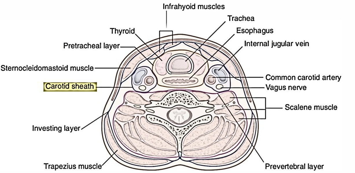
The carotid sheath goes from the base of the skull above to the arch of the aorta below. At the upper end it’s connected to the margins of carotid canal and the jugular fossa.
The upper part of the carotid sheath includes internal carotid artery, internal jugular vein and last 4 cranial nerves. Medial to it is located pharynx, lateral to it is located styloid equipment, anterior to it is located infratemporal fossa and posterior to it is located cervical sympathetic trunk on the prevertebral fascia.
The lower part of carotid sheath includes common carotid artery, internal jugular vein and vagus nerve.
Styloid Gear
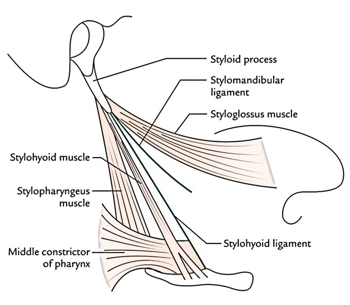
The styloid equipment is composed of styloid process and structures connected to it.
The structures connected to styloid process are:
- 3 muscles: stylohyoid, styloglossus and stylopharyngeus.
- 2 ligaments: stylohyoid and stylomandibular.
The 5 elongated structures connected to styloid process resemble the reins of a chariot. 2 of these reins (ligaments) are nonflexible, while the remaining 3 reins (muscles) are flexible, with every being controlled by individual cranial nerve, example, stylohyoid, stylopharyngeus and styloglossus are supplied by 7th, 9th and 12th cranial nerves, respectively.


 (53 votes, average: 4.58 out of 5)