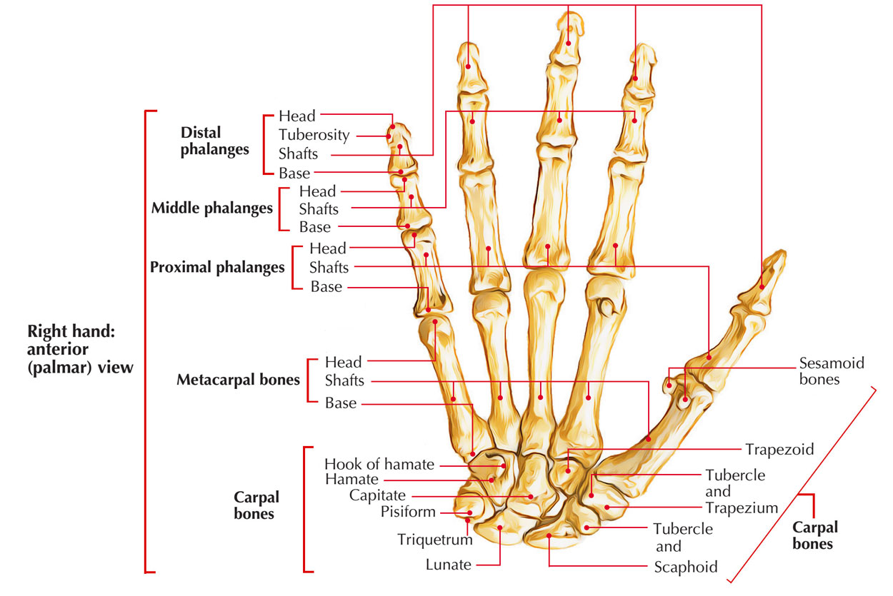
There are 3 groups of bones in the hand:
- The 8 carpal bones are the bones of the wrist.
- The 5 metacarpals (I to V) are the bones of the metacarpus.
- The phalanges are the bones of the digits -The thumb has only 2 and The remaining digits have 3.

Bones of the Hand
Carpal Bones

Carpal Bones
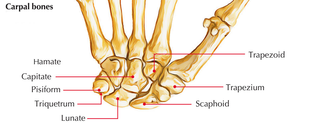
The carpus (G. Corpus = wrist) is composed of 8 carpal bones, that are arranged in 2 rows: proximal and distal. Every row contains 4 bones. The proximal row of carpal bones is composed of the following bones from lateral to medial side:
- Scaphoid
- Lunate
- Triquetral
- Pisiform
The distal row of carpal bones is composed of the following bones from lateral to medial side:
- Trapezium
- Trapezoid
- Capitate
- Hamate
Mnemonic: She Looks Too Pretty. Try To Catch Her.
Identification of Individual Carpal Bones
The individual carpal bones can be recognized by taking a look at their shape and few other features.
Identification of the carpal bones.
| Carpal bone | Identifying features |
|---|---|
| 1. Scaphoid | 1.Boat-shaped |
| 2.Has constriction (neck) | |
| 3.Has tubercle on distal part of its palmar surface | |
| 2. Lunate | Moon-shaped/crescentic |
| 3. Triquetral | 1.Pyramidal in shape |
| 2.Oval facet on the distal part of its palmar surface for articulation with pisiform | |
| 4. Pisiform | 1.Pea-shaped/pea-like |
| 2.Oval facet on the proximal part of its dorsal surface | |
| 5. Trapezium | 1.Quadrilateral in shape |
| 2.Has groove and crest (tubercle) on its palmar surface | |
| 6. Trapezoid | Shoe-shaped |
| 7. Capitate | 1.Largest carpal bone |
| 2.Has rounded head on its proximal surface | |
| 8. Hamate | 1.Wedge-shaped |
| 2.Hook-like process projects from distal part of its palmar surface |
Ossification
The carpal bones are cartilaginous at birth. Every carpal bone ossifies by 1 center and all these hearts seem after beginning. The centers seem as follows:
| Bones | Time |
|---|---|
| Capitate | Second month |
| Hamate | End of third month |
| Triquetral | Third year |
| Lunate | Fourth year in females, Fifth year in males |
| Scaphoid | Twelfth year in males, 9th to 10th in females |
Clinical Significance
Scaphoid fracture: Fracture of scaphoid is the most frequent fracture of carpus and normally happens because of fall on the outstretched hand. Fracture happens at the narrow waist of the scaphoid. Medically it presents as tenderness in the anatomical box. Blood vessels largely goes into the scaphoid via its both ends. But in 10-15% cases, all the blood vessels providing proximal section goes into it via its distal post. In this state when waistline of scaphoid is fractured, the proximal section is deprived of blood supply and may go through avascular necrosis.
Metacarpal Bones
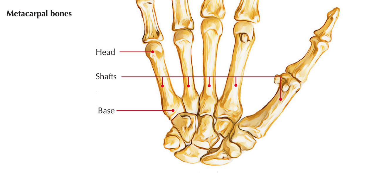
The metacarpus is composed of 5 metacarpal bones. They’re conventionally numbered 1 to 5 from lateral (radial) to medial (ulnar) side.

Metacarpal Bones
Parts
Every metacarpal is a small long bone and is composed of 3 parts: (a) head, (b) shaft, and (c) base.
Head
The head is at distal end and rounded.
Shaft
The shaft goes between head and base. It’s concave on palmar aspect and on sides. The dorsal surface of shaft presents a triangular area in its distal part.
Base
The base is proximal end and enlarged.
Peculiarities Of First Metacarpal
- The first metacarpal is the shortest and stoutest bone.
- It’s rotated medially via 90° so that its dorsal surface faces laterally.
- Its base possesses concavo-convex (saddle-shaped) articular surface for articulation with trapezium.
- The head is not as convex and wider than other metacarpals.
- The sesamoid bones glide on radial and ulnar corners of head and creates impressions of gliding.
- Its base dose not joint with any other metacarpal.
- It’s epiphysis at its proximal end contrary to other metacarpals, which have epiphysis at their distal end.
Ossification
Every metacarpal ossifies by 2 centers: 1 primary center for the shaft and the 1 secondary center for the head. The period of appearance of centers and their fusion is provided in the box below:
| Center | Time of appearance | Fusion |
|---|---|---|
| Primary centre for shaft | 9th week of IUL | |
| Secondary centre for head of second, third, fourth, and fifth metacarpal | 2 years | 16 years |
| Secondary centre for base for first metacarpal | 2 years | 18 years |
Clinical Significance
- Bennet’s fracture: It’s an oblique fracture of the base of 1st metacarpal. It’s intra-articular and could be related to subluxation or dislocation of metacarpal.
- Boxer’s fracture: It’s fracture of neck of metacarpal, and most generally includes neck of 5th metacarpal.
Phalanges
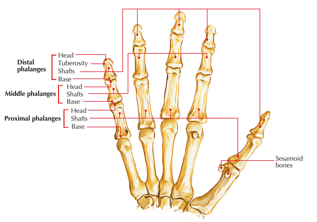
There are 14 phalanges in every hand: 2 in thumb and 3 in every finger.

Phalanges
Parts And Features
Every phalanx is a short long bone and has 3 parts: (a) base (proximal end), (b) head (distal end), and (c) shaft (going between the 2 ends).
Base
- The bases of proximal phalanges have concave oval facet for articulation with the heads of metacarpals.
- The bases of middle and distal phalanges possess pulley shaped articular surfaces.
Shaft
- The shaft tapers in the direction of the head.
- The dorsal surface is convex from side to side.
- The palmar surface is flat from side to side but gradually concave in the long axis.
Head
- The heads of proximal and middle phalanges are pulley shaped.
- The heads of distal phalanges is non-articular and has rough horseshoe-shaped tuberosity.
Ossification
Every phalanx ossifies by the 2 centers: 1 primary center for the shaft and 1 secondary center for the base. Their time of appearance is as follows:
Primary Centers
- For proximal phalanx: 10th week of IUL.
- For middle phalanx: 12th week of IUL.
- For distal phalanx: 8th week of IUL.
Secondary Centers
Look: 2 years. Fusion: 16 years.
Clinical Correlation
An undisplaced fracture of phalanx can be treated satisfactorily by strapping the fractured finger with the neighboring finger. The sesamoid bones in region of hand are found on the following sites:
- Sesamoid bone in the tendon of flexor carpi ulnaris (pisiform).
- Two sesamoid bones on the palmar surface of the head of first metacarpal.
- Sesamoid bone in the capsule of interphalangeal (IP) joint of thumb (in 75% cases).
- Sesamoid bone on the ulnar side of capsule of MCP joint of little finger (in 75% cases).
The sesamoid bones related to head of the first metacarpal bones are generally noticed in X-ray of hand.

 (55 votes, average: 4.57 out of 5)