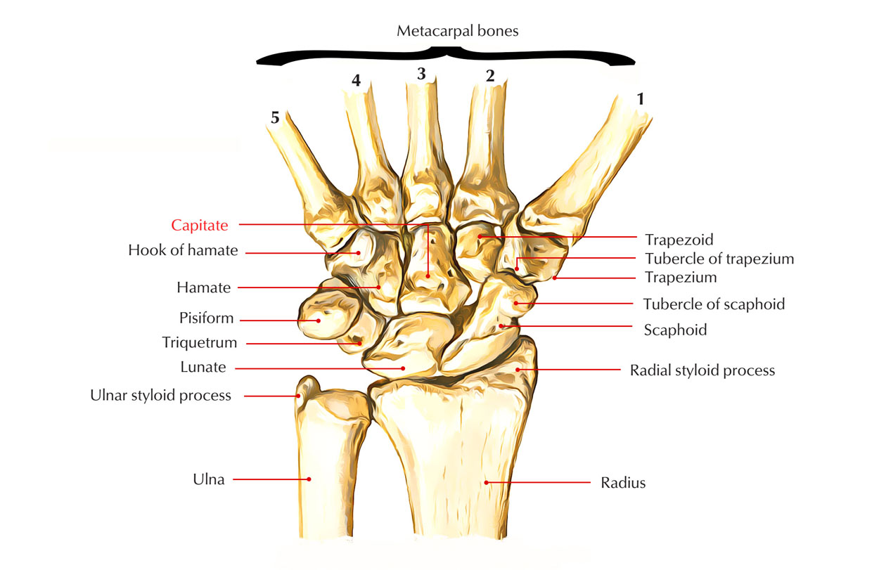
The capitate bone is the largest carpal bone located in the most central portion of the wrist. Distally the capitate bone articulates primarily along with the third metacarpal bone, however capitate bone likewise articulates along with the second and fourth metacarpal bones. Lateral element of capitate bone articulates along with the scaphoid (proximally) and along with the trapezoid (distally).
- Medially capitate bone articulates along with the hamate bone.
- The capitate bone lies right at the center of the carpus.
- Proximally, it articulates along with the lunate bone.
- The capitate bone bears a curved head at one end and is the biggest carpal bone.

Capitate Bone
Structure of Capitate Bone
The capitate bone looks like the scaphoid because the blood supply of capitate bone at the proximal pole is given by vessels flowing distally to the proximal, perhaps increasing the danger of fracture issues from disruption of the blood supply. The capitate is the biggest carpal bone.
- Distally, capitate bone articulates mainly along with the base of the third metacarpal and a small portion of the fourth.
- Radially along with the trapezoid.
- Proximally along with the scaphoid and lunate
- On its ulnar side along with the hamate
Surface of Capitate Bone
- The superior surface – round, smooth, and articulates along with the lunate bone
- The inferior surface – split by two rims into three sides for articulation along with the second, third, and fourth metacarpal bones, that for the third being the biggest
- The dorsal surface – wide and rough
- The palmar surface – narrow, rounded, and rough, for the connection of ligaments and a part of the adductor pollicis muscle
- The lateral surface – articulates along with the lesser multangular by a small side at its anterior inferior angle, behind which is a rough depression for the connection of an interosseous ligament. Superior to this is a deep, rough groove, forming part of the neck, and serving for the connection of ligaments; it is bounded superiorly by a smooth, convex surface, for articulation along with the scaphoid bone
- The medial surface – articulates along with the hamate bone by a smooth, concave, oval side, which inhabits its posterior and superior parts. It is rough in front, for the connection of an interosseous ligament.
Clinical Significance of Capitate Bone
Fractures
- Capitate bone fractures are categorized by the anatomic location of the fracture, together with what other concomitant injuries might exist.
- The force of injury in this syndrome can propagate resulting in perilunate dislocation too. The mix of a capitate bone fracture and a scaphoid waist fracture is called “scaphocapitate syndrome”.
- Different systems for fractures of the capitate bone have actually been postulated. The three systems-the first is direct trauma to the dorsal surface of the bone, the second is fall on the palm along with the wrist in forced extension and the third is fall on the powerfully bent hand; the second being the most regular and the third rarest.
- When it comes to a severe capitate bone fracture where there is x-ray proof of outstanding positioning of the fracture pieces, the attending physician will paralyze the wrist in a plaster or light-weight wrist brace. Once the cast has actually been eliminated the patient starts physiotherapy to gain back the variety of movement of the wrist joint and strength in the muscles included.
Treatment
Non-displaced capitate bone fractures have to be paralyzed just. Shift is a sign for fixation, as is concomitant surgery on the scaphoid; that is, if the scaphoid is fractured it will be corrected, and at that fixing the capitate fracture might be fixed too. Surgical fixation is included with a K-wire or a screw, along with a bone graft for considerable comminution.

 (54 votes, average: 4.82 out of 5)