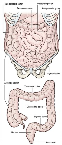
The Colon is part of the final stages of digestion. It split into 4 parts: (a) ascending colon, (b) transverse colon, (c) descending colon, and (d) sigmoid colon.
Ascending Colon
- The ascending colon is an upward continuation of the caecum. It’s about 5 inch (12.5 cm) in length and stretches from the caecum, at the level of ileocaecal orifice, to the inferior surface of the right lobe of the liver where it bends to the left to create the hepatic (right colic) flexure.
- It is located in the right paracolic gutter and covered by the peritoneum on the front and sides, which binds it to the posterior abdominal wall.
- Its posterior surface is located on 3 muscles: iliacus, quadratus lumborum, and transversus abdominis.
- During its course from the caecum to the undersurface of the liver, it crosses 3 nerves. From below upward these are: lateral cutaneous nerve of thigh, ilioinguinal nerve, and iliohypogastric nerve.
- Anteriorly it’s related to the coils of the small bowel and right edge of the greater omentum.
Points to be noticed
- Occasionally ascending colon possesses a mesentery named ascending mesocolon.
- Right margin of the greater omentum is occasionally fused with its peritoneal covering. Iff that’s the case, the right half of the transverse colon is frequently closely linked to the ascending colon, creating the so called” double barreled firearm”.
Transverse Colon
- It’s the longest (20 inch/50 cm in length) and most mobile part of the large intestine.
- It stretches from the right colic flexure (in right lumbar region) to the left colic flexure (in the left hypochondriac region).
- Strictly speaking transverse colon isn’t transverse but creates a dependent loop in front of loops of small intestine between the left and right colic flexures. The lowest point of loop generally goes up to the level of umbilicus but might occasionally extend into the pelvis. Therefore, the transverse colon is generally ‘U’ shaped.
Differences Between The Right (Hepatic) and Left (Splenic) Flexures
| Features | Right colic flexure | Left colic flexure |
|---|---|---|
| Location | Right lumbar region (where it is related to the inferior surface of right lobe of the liver) | Left hypochondrium (immediately below the spleen) |
| Level | 1 inch (2.5 cm) below the transpyloric plane, at the level of L2 vertebra | 1 inch (2.5 cm) above the transpyloric plane, at the level of T12 vertebra |
| Angulation | Wider | Acute |
| Attachment to diaphragm | No attachment | Attached to diaphragm at the level of 10th and 11th ribs by phrenicocolic ligament |
Relations
- Anterior: Greater omentum and anterior abdominal wall.
- Posterior: 2Nd part of the duodenum, head of the pancreas, and coils of the jejunum and ileum.
Clinical Significance
- The anterior surface of the transverse colon is closely linked to the anterior abdominal wall. This makes vulnerability of transverse colon simple for surgeons to do colostomy (making a hole in it).
- If the loops of the jejunum and ileum are distended, the transverse colon might be pushed up. Sometimes it enters anterior to the stomach and if distended with gas it might conceal the dullness of the liver to percussion and mimic the presence of gas in the peritoneal cavity.
Transverse Mesocolon
The transverse colon is suspended from the posterior abdominal wall by a large double-layered fold of the peritoneum referred to as transverse mesocolon.
- It’s fused with the posterior surface of the greater omentum and splits the peritoneal cavity into supra and infracolic compartments.
- Therefore, it creates a natural barrier against the spread of the infection between the supra and infracolic compartments.
- On the posterior abdominal wall, it’s connected to the 2nd part of the duodenum, head, and body (lower margin) of the pancreas and anterior surface of the left kidney.
- The contents of the transverse mesocolon are middle colic vessels, ascending branches of the left and right colic vessels, lymph nodes, lymphatics, and nerve plexuses embedded in the loose areolar tissue including varying amount of the fat.
Development
The proximal two-third of transverse colon grows from the midgut and the distal one-third from the hindgut.
Differences between The Right Two-Third and Left One-third of The Transverse Colon
| Features | Right two-third of transverse colon | Left one-third of transverse colon |
|---|---|---|
| Development | From midgut | From hindgut |
| Arterial supply | Middle colic artery, a branch of superior mesenteric artery (artery of midgut) | Left colic artery, a branch of inferior mesenteric artery (artery of hindgut) |
| Nerve supply | By vagus nerves | By pelvic splanchnic nerves |
Descending Colon
The descending colon is longer (25 cm), narrower, and more deeply found than the ascending colon. It goes from the left colic flexure to the very front of the left external iliac artery in the level of pelvic brim where it becomes continuous with the pelvic colon (sigmoid colon).
It’s covered by the peritoneum on the front and sides which fixes it in the left paracolic gutter and iliac fossa.
Course and Relationships
- Its proximal part descends vertically downward from the left colic flexure to the left iliac fossa. In this course it enters in front of 3 muscles and 3 nerves. The muscles are quadratus lumborum, transversus abdominis, and iliacus. The nerves are iliohypogastric, ilioinguinal, and lateral cutaneous nerve of the thigh.
- Its distal part turns medially from the left iliac fossa to the very front of the left external iliac vessels. In this course it enters in front of the femoral nerve, psoas major muscle, testicular vessels, genitofemoral nerve, and left external iliac vein.
Clinical Significance
The filled (with feces) descending colon may compress the left testicular and common iliac veins. This is one of the predisposing variables for greater frequency of varicosity of veins creating pampiniform plexus in the left spermatic cord (varicocele) and veins draining the left lower limb (varicose vein in the left lower limb).
Sigmoid Colon (Pelvic Colon)
The sigmoid colon is around 15 inches (37.5 cm) long and attaches the descending colon with the rectum. It’s S shaped and therefore its name, sigmoid colon (G. Sigma = S-shaped alphabet).
It goes from the lower end of descending colon in the left pelvic inlet to the pelvic surface of the 3rd section of sacrum, where it becomes continuous with the rectum.
During its course it creates a sinuous loop which hangs free in the lesser pelvis. In the pelvis it is located in front of the bladder and uterus, below the loops of ileum.
The loop of sigmoid colon contains 3 parts: (a) first part runs downward in contact together with the left pelvic wall; (b) 2nd part transverses the pelvic cavity horizontallybetween the bladder and the rectum in male (uterus and rectum in female); and (c) third part runs backward to get to the midline in front of third sacral vertebra.
Sigmoid (Pelvic) Mesocolon
The sigmoid colon is suspended from the pelvic wall by a large peritoneal fold termed sigmoid mesocolon. The sigmoid mesocolon has an inverted V shaped connection/ root.
The left limb: The left limb of the root is connected on the external iliac artery. It goes from the end of the descending colon to the middle of the common iliac artery. Here it turns sharply downward and to the right across the lesser pelvis to the 3rd section of the sacrum, creating the right limb. The right limb is connected on the pelvic outermost layer of the sacrum. The meeting point of 2 limbs is termed apex. The people must remember these facts in connection to the apex of “A”.
- Just lateral to the apex of the A, a pocket-like expansion of the peritoneal cavity enters upward posterior to the root of the mesocolon. It’s termed intersigmoid recess. The left ureter is located behind this recess.
- The inferior mesenteric artery splits near the apex of A.
- The superior rectal artery enters the right limb and sigmoidal arteries goes into the left limb.
Arterial Supply
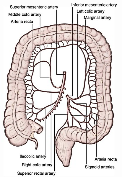
The colon is supplied by the following arteries:
- Ileocolic artery
- Right colic artery
- Middle colic artery
- Left colic artery
- Sigmoidal arteries.
- Superior rectal artery
The very first 3 arteries are the branches of the superior mesenteric artery and the past 3 are the branches of the inferior mesenteric artery.
The supply of distinct parts of the colon by these arteries is as under:
- The lower smaller part of the ascending colon is supplied by the ileocolic artery and its bigger upper part is supplied by the right colic artery.
- The right two-third of the transverse colon is supplied by the middle colic artery and the left one-third by the left colic artery.
- The descending colon is supplied by the left colic artery.
- The sigmoid colon is supplied by the sigmoidal branches of the inferior mesenteric artery and superior rectal artery. The sigmoidal arteries are generally 2 to 3 in number.
Marginal Artery of Drummond
- It’s a circumferential anastomotic arterial channel going from the ileocaecal junction to the rectosigmoid junction. It’s located close (about 3 cm) to the inner margin of the colon.
- It’s created by the anastomoses between the branches of colic branches of the superior mesenteric artery (i.e., ileocolic, right colic, and middle colic) and colic branches of the inferior mesenteric artery (left colic and sigmoidal). The vasa recta originate from the marginal artery and supply the colon.
Critical Point of Colon
It is located at the level of splenic flexure. At this level, the ascending branch of the left colic artery frequently neglects to anastomose with the left branch of the middle colic artery. Yet, there’s anastomotic channel between the principal trunk of the middle colic and the ascending branch of the left colic referred to as “arc of Riolan.” If this anastomosis isn’t well developed, the arterial supply of splenic flexure is endangered and splenic flexure goes through ischemic changes.
Before it was believed the critical point of the colon is really on the sigmoid colon (critical point of Sudeck) where the anastomosis between the sigmoidal branches of inferior mesenteric and superior rectal arteries was regarded as inadequate.
Venous Drainage
- The veins emptying the colon follow the arteries.
- The veins accompanying the ileocolic, right colic, and middle colic arteries join the superior mesenteric vein, while the veins, accompanying the branches of inferior mesenteric artery, join the inferior mesenteric vein. The superior and inferior mesenteric veins ultimately drain into the portal vein flow.
Lymphatic Drainage
The lymphatic drainage of the colon is medically very essential because carcinoma of the colon propagates via lymphatic route.
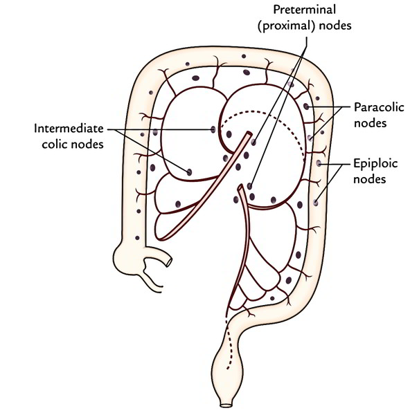
There are numerous colic lymph nodes, which drain the lymph from the colon. These nodes have common routine of distribution. They’re ordered in following 4 groups:
- Epiploic nodes, are small nodules and are located on the wall of the colon.
- Paracolic nodes, is located quite close to the marginal artery (of Drummond), i.e., along the medial edges of the ascending and descending colons and along the mesenteric edges of transverse and sigmoid colons.
- Intermediate colic nodes, is located along the ileocolic, right colic, middle colic and left colic, arteries, and drain into terminal nodes.
- Endmost nodes, is located along trunks of superior and inferior mesenteric arteries.
Clinical Significance
Evaluation of The Inside of Colon
Barium enema is utilized for visualizing the inner part of the colon. The normal routine of the colon because of the presence of sacculations is certainly observed. Today, endoscopic evaluation of the inside of the colon is commonly done.
Congenital Megacolon/Hirschsprung Disease
It happens when neural crest cells don’t migrate and create the myenteric plexus (parasympathetic ganglia) in the sigmoid colon and rectum during embryonic development. This state ends in absence of peristalsis. Consequently the normal proximal colon becomes grossly dilated because of the fecal retention causing abdominal distension. The constricted section normally corresponds to rectosigmoid junction.
Cancer (Carcinoma) of Colon
Cancer of colon (really large intestine) is a top cause of death in the Western world. It’s comparatively common in those who are above 50 years old and nonvegetarian. It’s slow growing tumor and causes constriction of the colon. The development is limited to the wall of colon for a substantial time before it propagates by lymphatics. In advanced cases, it spreads to the liver via portal vein circulation. If diagnosed early, hemicolectomy (partial resection of the colon) is carried out to heal the patient.
Surgical Resection For Cancer
While planning resection of the colon changed by carcinoma, the surgeon should remember the subsequent anatomical facts:
- Lymph from gut may miss the epiploic and paracolic nodes and go directly to the intermediate and preterminal nodes.
- The land of lymphatic drainage is split pretty correctly into regions corresponding to the regions supplied by the primary arteries.
- Removal of lymph nodes is possible only after ligating the principal arteries.
Therefore, the entire section of colon being supplied by the ligated artery ought to be taken out to prevent gangrene.
Sites of The Carcinoma In Colon
| Sites of carcinoma | Vessels to be ligated | Segments of colon to be removed |
|---|---|---|
| Caecum and ascending colon | Ileocolic, right colic, and right branch of middle colic | Terminal part of the ileum, caecum, ascending colon, hepatic flexure, proximal one-third of transverse colon |
| Hepatic flexure and right two-third of transverse colon | Ileocolic, right colic, and middle colic | Terminal part of the ileum, caecum, ascending colon, and right two-third of transverse colon |
| Left one-third of transverse colon and splenic flexure | Middle colic and left colic | From middle of the ascending colon to the beginning of sigmoid colon |
| Descending and sigmoid colons | Inferior mesenteric artery | Descending and pelvic colon |
Diverticulosis
The diverticulosis is a standard clinical condition of the colon and usually includes the sigmoid colon. It includes the herniation of the lining mucosa via the circular muscle between the teniae coli.
The herniation takes place where the circular muscle coat is the feeblest, i.e., where it’s pierced by the blood vessels.
The inflammation of diverticula is named diverticulitis.
- Volvulus: It’s a clinical illness, where a portion of gut rotates (clockwise/anticlockwise) on the axis of its mesentery. It typically happens because of adhesion of antimesenteric border of the gut to the parietes or some other viscera. It might correct itself spontaneously or the rotation may continue until the blood supply of the gut is cut off leading to ischemia. The sigmoid colon is susceptible to volvulus due to extreme freedom of its mesentery- the pelvic mesocolon.
- Intussusception: It’s a clinical condition where a proximal section of the bowel invaginates into the lumen of an adjoining distal section. This might cut off the blood supply to the bowel and cause gangrene. The different forms of intussusception are ileoileal, ileocaecal, and colocolic. The ileocaecal intussusception is the most typical form.


 (96 votes, average: 4.63 out of 5)