They can be called the main arteries of the head and neck. There are 2 common carotid arteries: left and right.
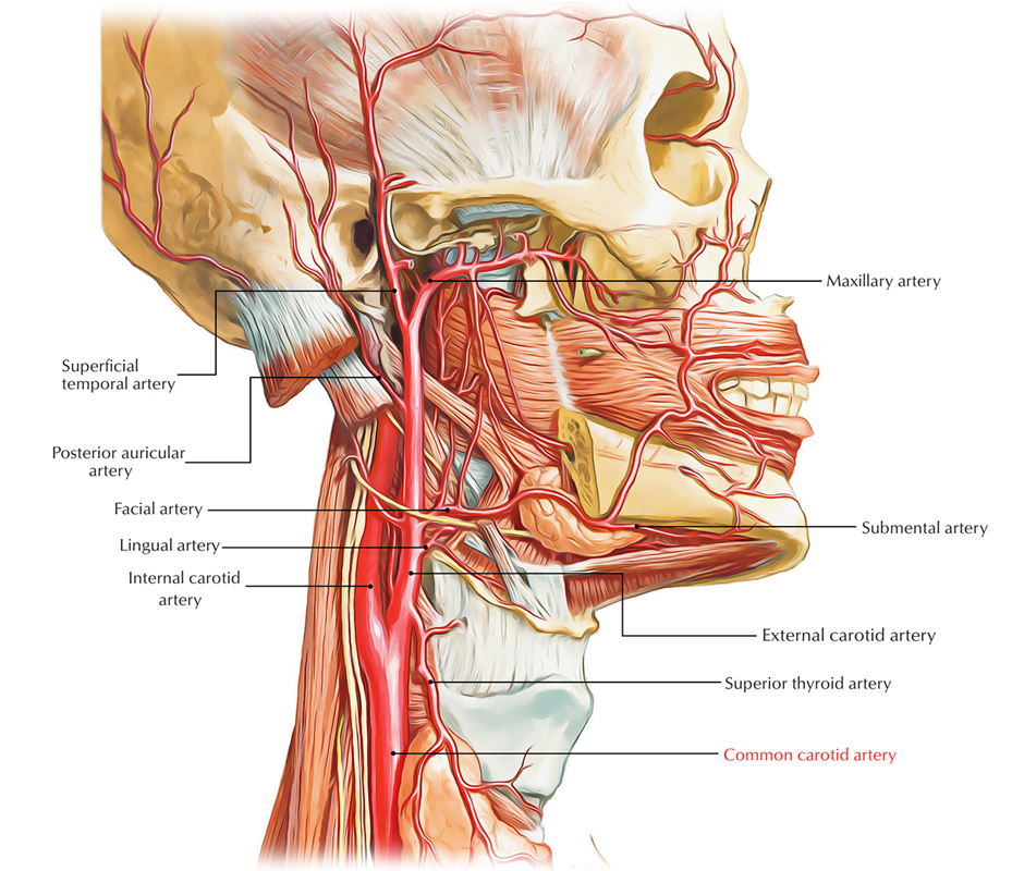
Common Carotid Artery
Origin
The right common carotid artery originates behind the sternoclavicular joint, in neck from brachiocephalic trunk (innominate artery).
The left common carotid artery originates directly from the arch of aorta in thorax (superior mediastinum). It ascends to the rear of left sternoclavicular joint and enters the neck.
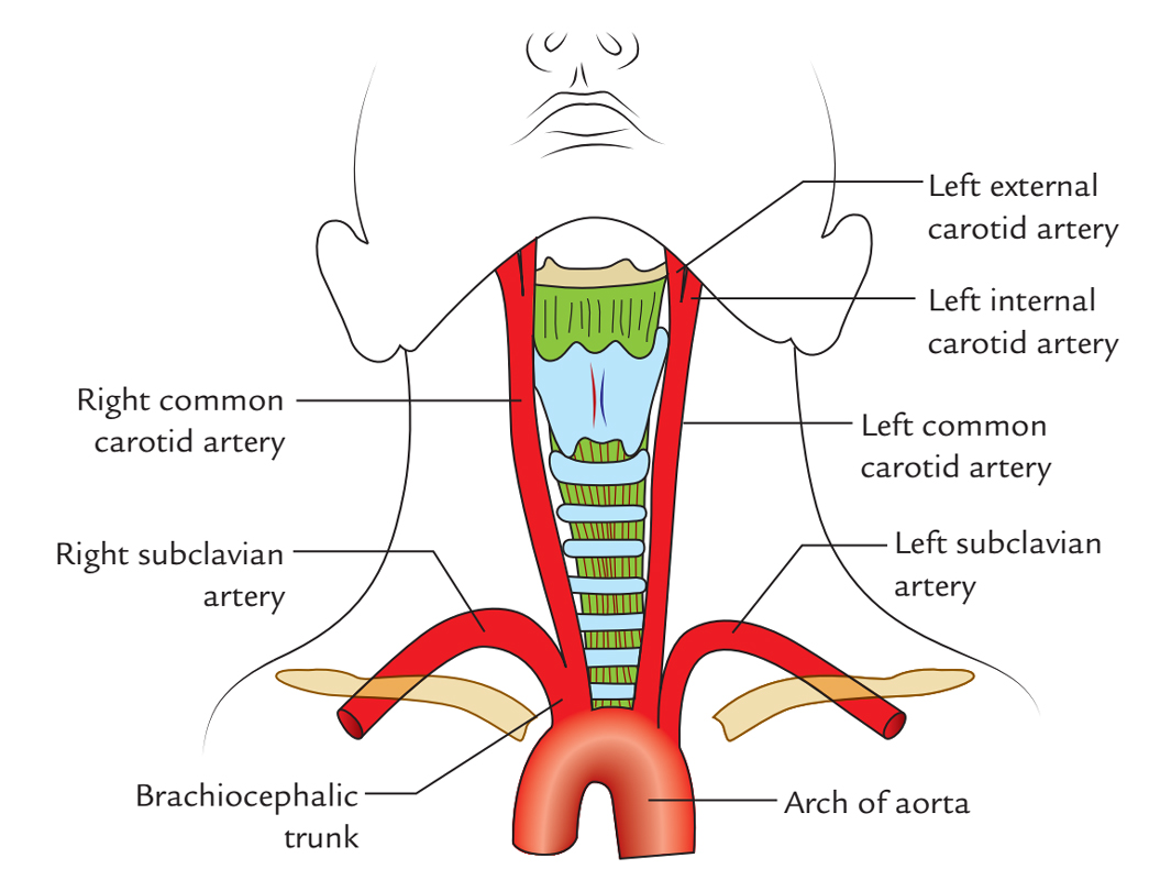
Common Carotid Artery: Origin
Course, Conclusion and Relationships
In the neck, both arteries (left and right) have quite similar course. Every artery runs upwards from sternoclavicular joint to the upper border of the lamina of thyroid cartilage (opposite the disk between the 3rd and 4th cervical vertebrae), where it ends by dividing into internal and external carotid arteries. The internal carotid artery is thought to be a continuance of common carotid artery. They’re referred to as internal and external because the former supplies structures inside the skull and latter supplies structures outside the skull.
Every common carotid artery is located in front of transverse processes of lower 4 cervical vertebrae under the cover of anterior border of the sternocleidomastoid muscle.
Branches
The common carotid artery supplies only 2 terminal branches, i.e., external and internal carotid arteries.
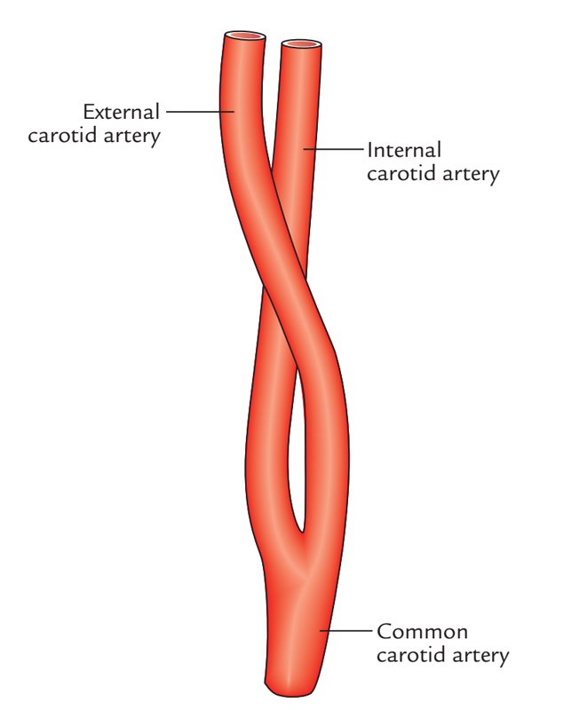
Common Carotid Artery: Branches
Clinical Significance Carotid Pulse
The common carotid artery can be compressed against the notable anterior tubercle of transverse process of the 6th cervical vertebrae referred to as carotid tubercle (Chassaignac’s tubercle) by pressing medially and posteriorly with the thumb. The carotid tubercle of the 6th cervical vertebra is situated about 4 cm above the sternoclavicular joint in the level of cricoid cartilage.
Above this level, the common carotid artery is superficial and therefore its pulsations can be easily felt. The carotid pulse is the steadiest pulse within the body.
External Carotid Artery
It’s 1 of the 2 terminal branches of the common carotid artery and supplies the structures external to the head and in front of the neck.
Course and Connections
The external carotid artery stretches upwards from the level of upper border of the lamina of the thyroid cartilage to a stage supporting the neck of the mandible, where it ends in the substance of the parotid gland by splitting into the superficial temporal and maxillary arteries.
The external carotid artery has a somewhat curved course so that it’s anteromedial to the internal carotid artery in its lower part and anterolateral to the internal carotid artery in its upper part.
Branches
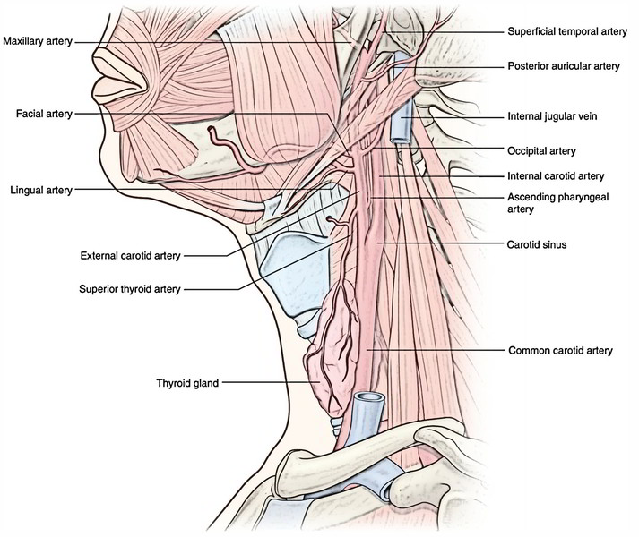
The external carotid artery supplies rise to 8 branches as follows:
- Superior thyroid artery.
- Lingual artery.
- Facial artery.
- Occipital artery.
- Posterior auricular artery.
- Ascending pharyngeal artery.
- Maxillary artery.
- Superficial temporal artery.
The very first 3 arteries originate from anterior aspect, next 2 from posterior aspect and next 1 from medial aspect. The past 2 are terminal branches.
The branches of the external carotid artery are 8 in number (Mnemonic the term EXTERNAL is composed of 8 letters: 1, 2, 3, 4, 5, 6, 7 and 8 which correspond to the number of branches of the external carotid artery).
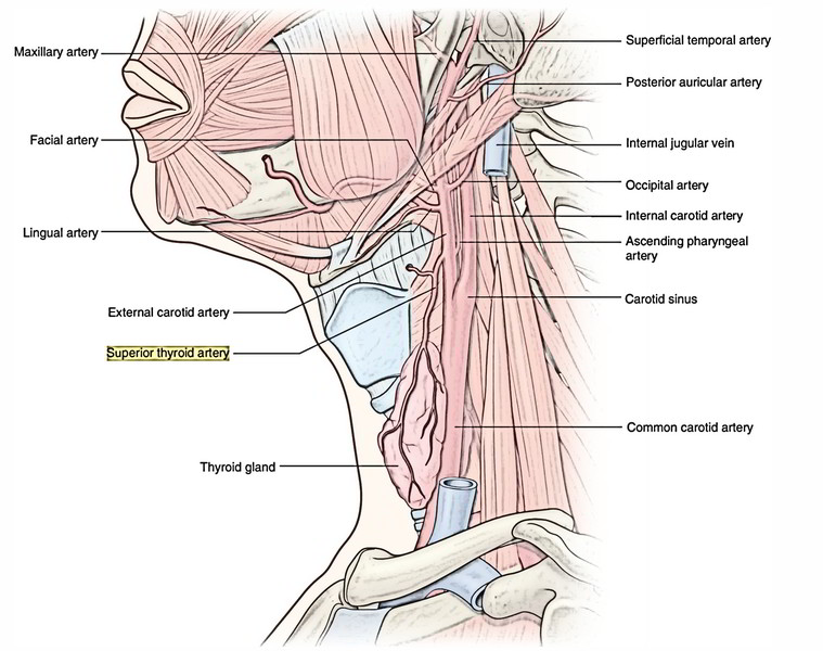
Superior Thyroid Artery
It appears from the front of external carotid artery just below the tip of the higher cornu of the hyoid bone. It runs downwards and forwards, parallel and superficial to the external laryngeal nerve to make it to the upper pole of the thyroid gland, which it furnishes.
Branches.
- Infrahyoid branch, which anastomoses with its fellow of opposite side.
- Sternomastoid branch to the sternomastoid muscle.
Superior laryngeal artery accompanies the internal laryngeal nerve, enters deep to the thyrohyoid muscle and pierces the thyrohyoid membrane to supply the larynx.
Cricothyroid branch, enters across the cricothyroid ligament and anastomoses with its counterpart of the opposite side.
Glandular branches to the thyroid gland; 1 of which anastomoses with its fellow of the opposite side along the upper border of the isthmus of the thyroid gland
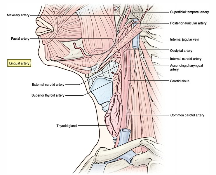
Lingual Artery
It appears from the very front of the external carotid artery opposite the tip of the higher cornu of the hyoid bone. It’s the primary artery to supply blood to the tongue. It might originate in common with the facial artery (linguofacial trunk).
It’s split into 3 parts by the hyoglossus muscle, viz.
First part is located in the carotid triangle and creates a feature loop with convexity upwards above the greater cornu. The loop is crossed superficially by the hypoglossal nerve. The loop allows free movement of the hyoid bone.
Second part is located deep to the hyoglossus muscle along the upper border of the hyoid bone.
Third part (also referred to as arteria profunda linguae) or deep lingual artery first runs upwards along the anterior border of the hyoglossus muscle and after that forwards on the undersurface of the tongue, where it anastomoses with its fellow of opposite side.
Branches
- From first part-suprahyoid branch. It anastomoses with its fellow of opposite side.
- From second part-dorsal linguae branches generally 2 in number, to the dorsum of tongue and tonsil.
- From third part-sublingual artery, to the sublingual gland
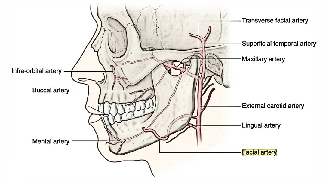
Facial Artery (Once Referred To As External Maxillary Artery)
It appears from the very front of the external carotid artery just above the tip of the higher cornu of the hyoid bone.
It’s split into 2 parts-cervical and facial:
- Cervical part of the facial artery ascends deep to the digastric and stylohyoid muscles, enters deep to the ramus of mandible where it grooves the posterior border of the submandibular gland Afterward it makes S shaped crook, first bending down (with convexity upwards) over the submandibular gland and after that upward (with convexity downwards) over the base of the mandible.
- Facial part of the facial artery starts where the facial artery winds around the lower border of the body of the mandible in the anteroinferior angle of the masseter.
Branches
From the cervical part (branches in the neck).
- Ascending palatine artery originates near the origin of facial artery, ascends and accompanies the levator palati, enters over the upper border of the superior constrictor and supplies primarily the palate.
- Tonsillar artery (principal artery of tonsil) pierces the superior constrictor and ends in the tonsil.
- Glandular branches supply the submandibular gland
- Submental artery, a large artery which runs forwards on the mylohyoid muscle in business with mylohyoid nerve. It supplies the mylohyoid muscle and submandibular and sublingual salivary glands.
Occipital Artery
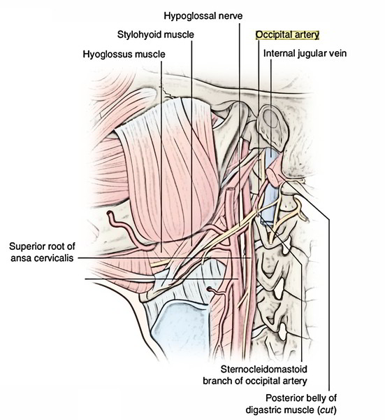
It originates from the posterior aspect of the external carotid artery at precisely the same level as the facial artery. It runs backwards and upwards under cover of lower border of the posterior belly of digastric muscle superficial to internal carotid artery, internal jugular vein and last 4 cranial nerves, crosses the apex of the posterior triangle. Afterward it runs deep to the mastoid process grooving the lower surface of the temporal bone medial to the mastoid notch. It crosses the superior oblique and semispinalis capitis and apex of suboccipital triangle to reach underneath the trapezius muscle, which it pierces 2.5 cm far from the midline and comes to is located just lateral to the greater occipital nerve. It supplies majority of the rear of the entire scalp.
Branches
Sternomastoid branches are generally 2 in number. They run downwards and backwards and supply the sternocleidomastoid. The upper 1 accompanies the spinal accessory nerve and lower 1 is hooked by the hypoglossal nerve.
- Mastoid branch enters the cranial cavity via mastoid foramen and supplies mastoid air cells.
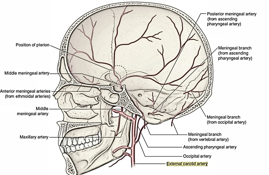
- Meningeal branches goes into the cranial cavity via jugular foramen and hypoglossal canal to supply dura mater of posterior cranial fossa.
- Muscular branches supply adjoining muscles.
- Auricular branch (occasional) supplies the cranial surface of the auricle.
Descending branch breaks up into superficial and deep branches. The superficial branch anastomoses with the superficial branch of transverse cervical artery and deep branch anastomoses with the deep cervical artery-a branch of the costocervical trunk of subclavian artery on the superficial and deep surfaces of the semispinalis capitis, respectively.
Clinical significance
The descending branch of the occipital artery gives the main collateral circulation after ligation of the external carotid or the subclavian artery (vide supra).
Key Points
- The hypoglossal nerve hooks the occipital artery under its site of origin.
- The upper sternomastoid branch of occipital artery accompanies the spinal accessory nerve and the lower sternomastoid branch crosses the hypoglossal nerve.
- Occipital artery crosses the apical part of the posterior triangle.
Posterior Auricular Artery
It appears from the posterior aspect of the external carotid artery a little above the occipital artery. It crosses superficial to the stylohyoid muscle. It runs upwards and backwards parallel to the occipital artery along the upper border of the posterior belly of digastric muscle and deep to the parotid gland. Subsequently it becomes superficial and is located on the base of mastoid process supporting the ear which it supplies.
Branches
- Stylomastoid artery enters the stylomastoid foramen to supply middle ear, mastoid air cells and facial nerve.
- Auricular branch supplies both cranial and lateral surfaces of the auricle.
- Occipital branch, supplies scalp above and behind the auricle.
Clinical significance
The posterior auricular artery is cut in incisions for mastoid procedures.
Ascending Pharyngeal Artery
It’s a slight artery that originates from the medial aspect of the external carotid artery near its lower end. It runs vertically upwards between the side wall of the pharynx and internal carotid artery up to the base of the skull.
Branches
- Pharyngeal and prevertebral branches to corresponding muscles.
- Meningeal branches, which traverse foramina in the base of the skull.
- Inferior tympanic, which supplies medial wall of tympanic cavity.
- Palatine branches, which follow levator veli palatini to the palate.
Superficial Temporal Artery
It’s the smaller but more direct terminal branch of the external carotid artery. It starts behind the neck of the mandible deep to the upper part of the parotid gland It runs vertically upwards crossing the root of zygoma in front of the tragus where its pulsation can be felt.
About 5 cm above the zygoma, it splits into anterior and posterior branches, which supply the temple and scalp.
Branches.
- Transverse facial artery runs forwards across the masseter below the zygomatic arch.
- Anterior auricular branch, supplies the lateral surface of auricle and external auditory meatus.
- Zygomatico-orbital artery runs forwards along the upper border of zygomatic arch between 2 layers of temporal fascia and reaches the lateral angle of the eye.
- Middle (deep) temporal artery runs on the temporal fossa deep to temporalis muscle and supplies temporalis muscle and fascia.
- Anterior (frontal) and posterior (parietal) terminal branches.
The anterior branch supplies the muscles and skin of the frontal region. It’s quite tortuous and anastomoses with the branches of the ophthalmic artery. The posterior branch sup-plies skin and the auricular muscles.
Clinical significance
Superficial temporal pulse: The pulsations of superficial temporal artery can be easily felt in front of the tragus of the ear (where it crosses the root of zygoma, the preauricular stage). It serves the useful function to anesthetists to whom the radial pulse isn’t accessible during surgery. For this particular reason, it’s also termed anesthetist’s artery.
The course of anterior terminal branch of the superficial temporal artery on the brow can certainly be viewed in a hairless furious guy. It becomes visibly more tortuous with increasing age.
Maxillary artery: It’s the bigger terminal branch of the external carotid artery.
Internal Carotid Artery
The internal carotid artery is 1 of the 2 terminal branches of the common carotid artery but it’s more direct. It’s regarded as an upward continuation of the common carotid artery. It supplies structures within the skull and in the orbit. It’s the main artery to supply the brain and eye.
It starts as the upper border of the lamina of thyroid cartilage in the level of the disk between C3 and C4 vertebrae and runs upwards to get to the base of the skull, where it enters the carotid canal in the petrous temporal bone. It comes in the cranial cavity by going through the upper part of the foramen lacerum. In the cranial cavity, it enters the cavernous sinus and pursues a tortuous course before it finishes below the anterior perforated substance of the brain by splitting into the anterior and middle cerebral arteries.
Parts
For the interest of convenience, the course of the internal carotid artery is split into the following 4 parts:
Cervical Part
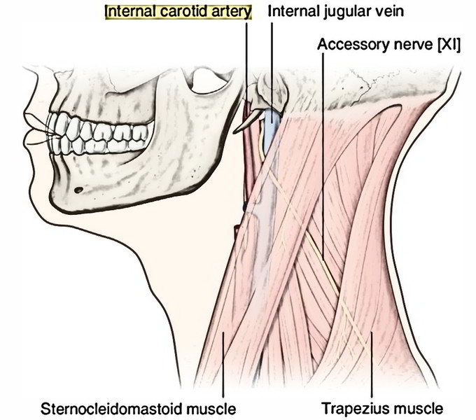
It ascends vertically upwards from its ori-gin to the base of the skull to reach the lower end of carotid canal and is located on the front of transverse process of upper cervical vertebrae. In the neck, the internal carotid artery is enclosed in the carotid sheath together with the internal jugular vein and vagus nerve.
The lower part of the artery is superficial and located in the carotid triangle. The upper part is profoundly locatedand is located deep to the posterior belly of digastric muscle, styloid process with structures connected to it and parotid gland
At the upper end, the internal jugular vein is located posterior to the internal carotid artery. Here the last 4 cranial nerves (glossopharyngeal, vagus, accessory and hypoglossal) is located between the internal jugular vein and internal carotid artery.
Branches
In the neck, the internal carotid artery supplies no branches.
Petrous Part
The internal carotid artery enters the petrous part of the temporal bone in a carotid canal. It first runs upwards and after that turns forwards and medially at the right angle. It appears at the apex of petrous temporal bone in the posterior wall of foramen lacerum and goes through its upper part to goes into the cranial cavity.
Branches
- Caroticotympanic branches to middle ear, which anastomose with the anterior and posterior tympanic arteries.
- Pterygoid branch (small and inconstant) enters the pterygoid canal and anastomoses with the greater palatine artery.
Cavernous Part
This part is located inside the cavernous sinus. From foramen lacerum, the internal carotid artery ascends and enters the cavernous sinus. In the sinus, it enters forwards along the side of sella turcica in the floor and medial wall of the sinus. Here it is located outside the endothelial lining of the sinus and related to the abducent nerve inferolaterally.
In the anterior part of the sinus, the artery ascends and pierces the dural roof of the sinus between the anterior and middle clinoid processes to reach underneath the cerebrum.
Branches
- Cavernous branches to the trigeminal ganglion.
- Superior and inferior hypophyseal arteries to the hypophysis cerebri (pituitary gland).
Cerebral Part
This part is located at the base of the brain. After appearing from the roof of the cavernous sinus, the artery turns backwards in the subarachnoid space along the roof of the cavernous sinus and is located below the optic nerve.
Eventually it turns upwards by the side of the optic chiasma and reaches the anterior perforated substance of the brain found at the start of the stalk of lateral sulcus of the cerebral hemisphere. Here it ends by breaking up into anterior and middle cerebral arteries.
Outline of Branches of the Internal Carotid Artery
| Part | Branches |
|---|---|
| Cervical part | No branches |
| Petrous part | Caroticotympanic branches Pterygoid branch |
| Cavernous part | Cavernous branches Superior and inferior hypophyseal arteries |
| Cerebral part | Ophthalmic artery Anterior choroidal artery Posterior communicating artery Terminal branches Anterior cerebral artery Middle cerebral artery |
Carotid siphon: The U shaped curve created by the internal carotid artery while going through and above the cavernous sinus is known as carotid siphon. It likely dampens down the pulsations of the artery. The carotid siphon is a significant characteristic to be observed in cerebral angiography.
Clinical significance
The carotid vessels are exposed during optional arterial surgery for aneurysms, arteriovenous fistulae, or arteriosclerotic occlusion. It’s now confirmed that partial or complete blockage because of arteriosclerosis is a familiar cause of cerebral stroke. Actually, it might account for 20% of the cerebrovascular accidents, which at one time were considered to be either because of intracranial hemorrhage or thrombosis.
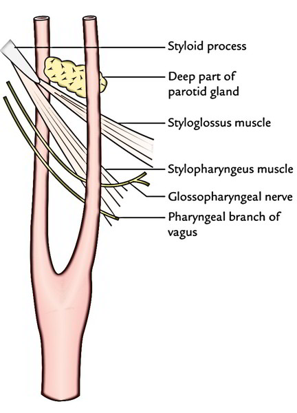
The structures passing between the external and internalcarotid arteries are as follows:
- Deep part of the parotid gland
- Styloid process.
- Styloglossus muscle.
- Stylopharyngeus muscle.
- Glossopharyngeal nerve.
- Pharyngeal branch of the vagus nerve.

 (63 votes, average: 4.65 out of 5)