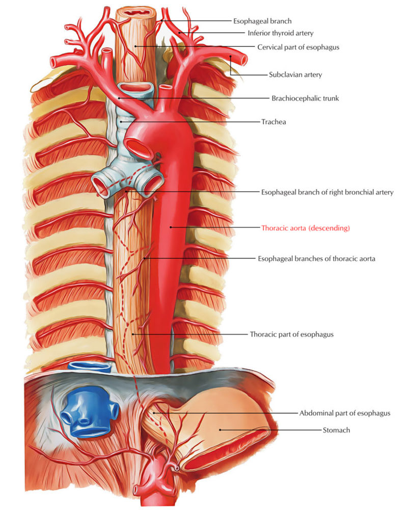
The descending aorta is the section of the thoracic aorta which is constrained into the posterior mediastinum. It originates leveled along with the lower boundary of the fourth thoracic vertebra, consistent with the aortic arch, and also terminates anterior to the lower boundary of the twelfth thoracic vertebra within the aortic hiatus. As it goes down, it enters as well as ends within the midline but on its genesis, it is left of the spinal column.
Structure

Descending Aorta
The majority of the descending aorta is created through the combination of the primitive dorsal aortae. In embryos around 2-6 mm. long, having twenty-three mesodermal somites, the primitive dorsal aorta; are merged with each other through the tenth to the sixteenth section. On a later stage the fusion is carried on backwards towards the twenty-third body section the level of the fourth lumbar vertebra exactly where the common iliac arteries emerge. Still later the small terminal sections of the primitive dorsal aorta merge with each other to create the unpaired middle sacral artery, which ends on the termination of the coccygeal region.
Relations
- Anterior to the descending thoracic aorta, from superior to inferior, is the left pulmonary hilum.
- The pericardium dividing it via the left atrium, esophagus along with the vertebral part of the diaphragm.
- The vertebral column as well as hemiazygos veins are posterior.
- Posterior towards the right are the azygos vein along with thoracic duct, and inferiorly, the right pleura and lung.
- The pleura as well as lung are towards the left.
Surface Anatomy
The descending thoracic aorta might be protruded as a 2.5 cm wide strap via the sternal termination of the second left costal cartilage into a median placement 2 cm over the trans-pyloric plane on the first lumbar vertebra.
Position
Even though the descending aorta is located towards the left of the body’s midline, it progressively goes down towards immediately facing the vertebral column on the left of the 12th thoracic vertebra. The part of the descending aorta over the diaphragm is referred to as the thoracic aorta, and also produces sections within the thoracic wall. These sections, the bronchial, pericardial, as well as esophageal arteries, circulate blood towards the organs for which they were entitled. The esophagus along with its plexus of nerves lies sideways as well as towards the right within the upper thorax, anteriorly within the lower thorax and also comes down towards a left anterolateral position when it enters the diaphragm. Up to a certain level, the descending aorta as well as esophagus is jointly spiraled. Sometimes, the aorta goes into the diaphragm towards the left as well as at the back of the esophagus.
Branches
It possesses six paired sections: bronchial arteries, mediastinal arteries, esophageal arteries, pericardial arteries, superior phrenic artery, and intercostal arteries. There are nine pairs of the intercostal arteries. The right sections are lengthier compared to the left, since the descending aorta (thoracic aorta) is at the left portion of the vertebrae. Via its numerous branches, it circulates blood towards the esophagus, lungs, as well as the chest region, consisting of the ribs along with mammary glands. The thoracic aorta gives visceral sections towards the pericardium, lungs, bronchi along with oesophagus, as well as parietal divisions toward the thoracic wall.

 (48 votes, average: 4.85 out of 5)