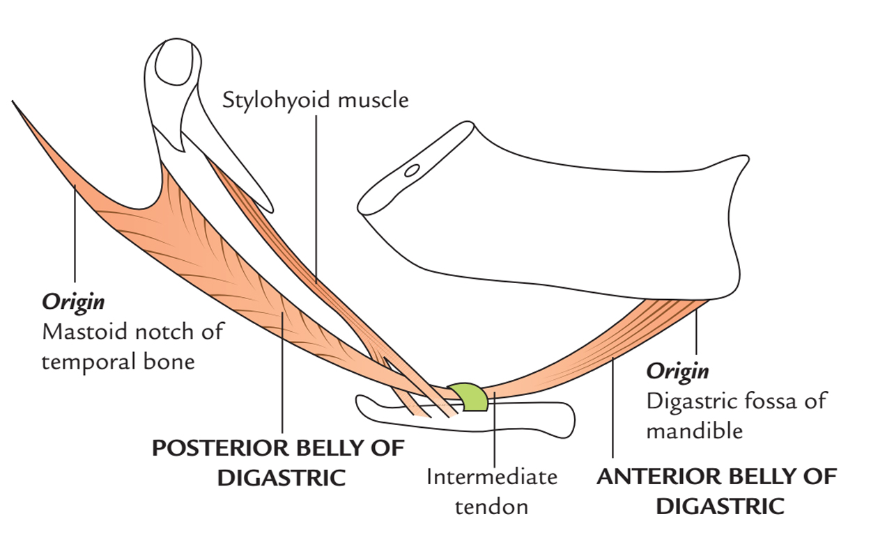
Digastric muscle is a muscular band that is made up of posterior and anterior bellies, which are joined by an intermediate tendon. Because of its two bellies this muscle is named digastric.

Digastric Muscle
Origin
- The bipinnate posterior belly- via the mastoid notch of the temporal bone.
- The unipinnate anterior belly – via the digastric fossa on the lower margin of the mandible near the symphysis menti.
Insertion

Digastric Muscle: Origin and Insertion
Both anterior and posterior bellies are attached in the intermediate tendon. The intermediate tendon is attached by an upside-down U-shaped facial sling of enclosing layer of deep cervical fascia to the junction of the body as well as greater cornu of hyoid bone. The tendon of digastric muscle travels in the middle of the two strips of tendon of stylohyoid muscle.
Structure
Digastric muscle has two bellies anterior and posterior. The embryological origins of two bellies of the digastric muscle are not the same. The digastric muscle spreads out from the mastoid process of the cranium to the chin at mandible. In the midway it transforms into a tendon that travels via a tendinous pulley and attaches with the hyoid bone.
Posterior belly
- Compared to the anterior belly, the posterior belly is longer and originates via the mastoid notch which is located medial towards the mastoid process of the temporal bone, on the inferior side of the skull.
- The mastoid notch is a deep indentation in the middle of the mastoid process and the styloid process.
- The mastoid notch is also known as the digastric groove or the digastric fossa. The digastric branch of facial nerve innervates the posterior belly.
Anterior belly
- The anterior belly originates via a indentation on the inner side of the lower margin of the mandible referred to as the digastric fossa of mandible, which is located near the symphysis and goes inferior and posterior.
- The anterior body is innervated via the trigeminal nerve through the mylohyoid nerve.
- It arises via the first pharyngeal arch.
Intermediate tendon
The two bellies of digastric muscle terminate in an intermediate tendon that pierces the stylohyoideus muscle.
A fibrous loop, which is occasionally covered via a mucous sheath, it is held along the side of the body and the greater cornu of the hyoid bone.
Triangles
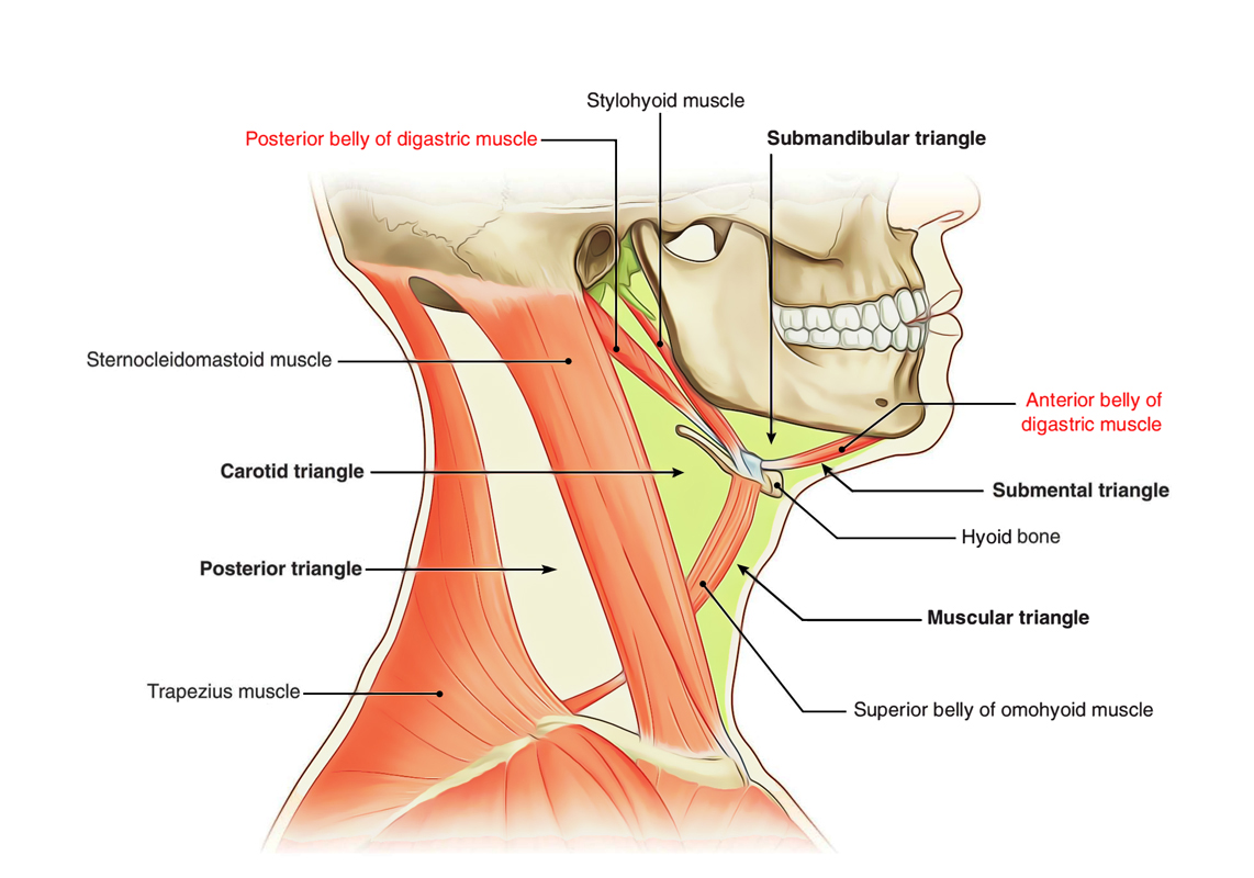
The Digastricus splits the anterior triangle of the neck within three smaller triangles:
- The submandibular triangle a.k.a. digastric triangle is surrounded:
- Superiorly via the lower margin of the body of the mandible along with a depicted line via mandibular angle towards the sternocleidomastoid.
- Inferiorly via the posterior belly of the digastricus as well as the stylohyoideus.
- Anteriorly via the anterior belly of the digastricus.
- The carotid triangle which is surrounded:
- Superiorly via the posterior belly of the digastricus and stylohyoideus.
- Posteriorly via the sternocleidomastoideus.
- Inferiorly via the omohyoideus.
- The suprahyoid a.k.a. submental triangle is surrounded:
- Laterally via the anterior belly of the digastricus.
- Medially via the middle line of the neck via the hyoid bone till the symphysis menti.
- Inferiorly via the body of the hyoid bone.
- The inferior carotid triangle a.k.a. muscular triangle is bounded:
- Anteriorly via the median line of the neck via the hyoid bone till the sternum.
- Posteriorly via the anterior border of the Sternocleidomastoideus.
- Superiorly via the superior belly of the omohyoideus.
Nerve Supply
- Posterior belly – Facial nerve.
- Anterior belly – Mylohyoid nerve.
Arterial Supply
- Anterior belly – Submental branch of the facial artery.
- Posterior belly – Occipital artery.
Actions
- Whenever the mouth is opened widely against opposition the digastric muscle helps in order to depress the mandible.
- Pulls the hyoid bone upwards at the time of deglutition.

 (61 votes, average: 4.70 out of 5)