Dorsal Interossei of Hand
Derivation and Terminology of Dorsal Interossei
The word “Dorsal”
- Indicates “the back” which is originated from the Latin – dorsalis or dorsum
- Dorsal usually is used to specify the back, or the “back side.”
The word “Interossei”
- The Latin inter, which indicates “between” or “among”
- Ossei means “bone.” which is originated from ossis,.
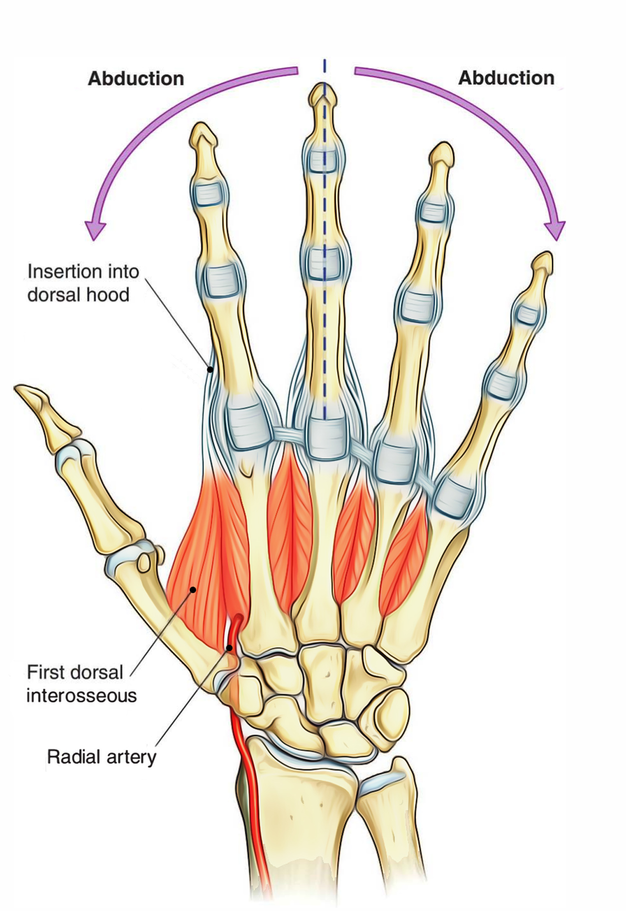
The dorsal interossei are the muscles between the bones and on the back side of the hand.

Dorsal Interossei (Hand)
Origin
There are four dorsal interossei:
- The first arises from adjacent sides of the thumb and index metacarpal
- the second from the surrounding sides of the index and long metacarpal
- the third from the surrounding sides of the long and ring metacarpals
- the fourth from the adjacent sides of the ring and small metacarpals
Insertion
- The first dorsal interosseous inserts into:
- the dorsal aponeurosis of the extensor hood of the index finger.
- and into the radial side of the bottom of the index proximal phalanx.
- The second enters into:
- The dorsal aponeurosis of the extensor hood of the long finger.
- And right into the radial side of the bottom of the long finger proximal phalanx.
- The third inserts into:
- The dorsal aponeurosis of the extensor hood of the long finger.
- And into the ulnar side of the base of the proximal phalanx of the long finger.
- The fourth enters into:
- the dorsal aponeurosis of the extensor hood of the ring finger.
- and within the ulnar side of the base of the proximal phalanx of the ring finger.
- The amount connecting within the extensor versus the associated proximal phalanx is not the same for each digit, in regards to the relative quantities of insertion.
- The first dorsal interosseous, the extensor hood inserts primarily into the proximal phalanx with a lesser part inserting into.
- Generally, the second and fourth have considerable contributions to both the associated proximal phalanx and to the dorsal aponeurosis, but, the second, third, and fourth have variable insertions.
- The third dorsal interosseous mainly enters into the dorsal aponeurosis of the long finger, along with a minimal component inserting into the base of the proximal phalanx
Nerve Supply
The deep branch of the ulnar nerve, root value T1 supplies all four dorsal interossei. The roots C6, C7 and C8 supply the skin overlying the muscles.
Vascular Supply
- Dorsal metacarpal arteries
- second to fourth palmar metacarpal arteries
- small branches of the radial artery
- arteria princeps pollicis
- arteria radialis indicis
- Perforating branches from the deep palmar arch (proximal perforating arteries)
- 3 distal perforating arteries
- dorsal digital arteries
Action
- The index, long, and ring finger, proximal phalanges, drawn away from the mid-axis of the long finger by the dorsal interossei.
- The MCP joints are also flexed by dorsal interossei muscles.
- The dorsal interossei help to extend the PIP and DIP joints, through the extensor hood.
- The functions of the interossei vary among the digits because each dorsal interosseous muscle varies in the relative amounts of entarnce within the proximal phalanx or into the dorsal aponeurosis.
Examination
- The index, middle or ring fingers the dorsal interossei can be examined on the dorsum of the hand in between the metacarpals during resisted abduction.
- In the thumb web, the first dorsal interosseous may be seen contracting in opposition to resistance.
- The palmar interossei and lumbricals are too deep to palpate but their activities can be demonstrated by accurate electrical stimulation.
Dorsal Interossei of Foot
The dorsal introsseous muscles exist on the lateral side only in third and fourth toes. In the first layer of muscles in the sole of the foot, the great and little toes have their own abductors (the abductor hallucis and abductor digiti minimi).
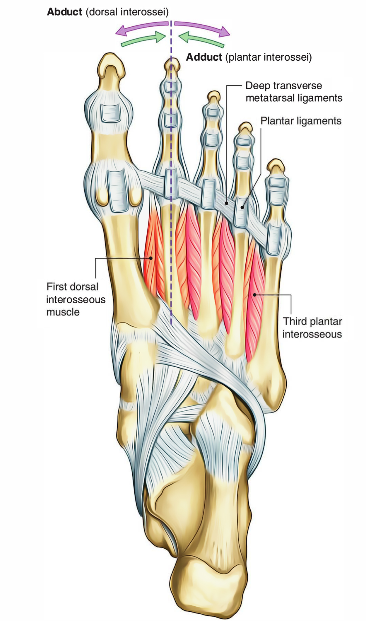
The four dorsal interossei abduct the second to fourth toes relative to the long axis via the second toe and are the most superior muscles in the sole of the foot.
All four muscles emerge from the sides of adjacent metatarsals and are bipennate. The tendons of the dorsal interossei insert into the base of the proximal phalanges of the toes and free margin of the extensor hoods.
The second toe has two dorsal interossei connected with it, one on each side, so it can be abducted to both side of its long axis.

Dorsal Interossei (Foot)
Origin
- The first, or most medial, attaches to the medial side of the base of the proximal phalanx of the second toe and arises from the adjacent sides of the first and second metatarsals.
- The second also attaches to the proximal phalanx of the second toe, but, to the lateral side and it arises from the adjacent sides of the second and third metatarsals.
- The third and fourth dorsal interossei attach to the lateral side of the proximal phalanx of the third and fourth toes respectively.
Nerve Supply
All four dorsal interossei are supplied by:
- the lateral plantar nerve, root value S2, S3
- those in the fourth interosseous space from the superficial branch, and the rest by the deep branch
- The skin covering this area on the dorsum of the foot is supplied by root L5 medially and S1 laterally
Action
- They produce flexion of the metatarsophalangeal joint, acting with the plantar interossei.
- The dorsal interossei abduct the toes at the metatarsophalangeal joint, however, this action, as such, is of little importance in the foot.
Functional Activity
- The dorsal interossei are powerful little muscles and their activity in combination with the plantar interossei controls the direction of the toes during forceful activity, thus enabling the long and short flexors to carry out their appropriate actions.
- These muscles, because of their relationship to the metatarsophalangeal joint.
- Are able to contract these joints, therefore, raise the heads of the second, third and fourth metatarsals,.
- Therefore helping to maintain the anterior metatarsal arch.
- They also help, to a limited extent with the maintenance of the medial and lateral longitudinal arches of the foot.

 (56 votes, average: 4.62 out of 5)