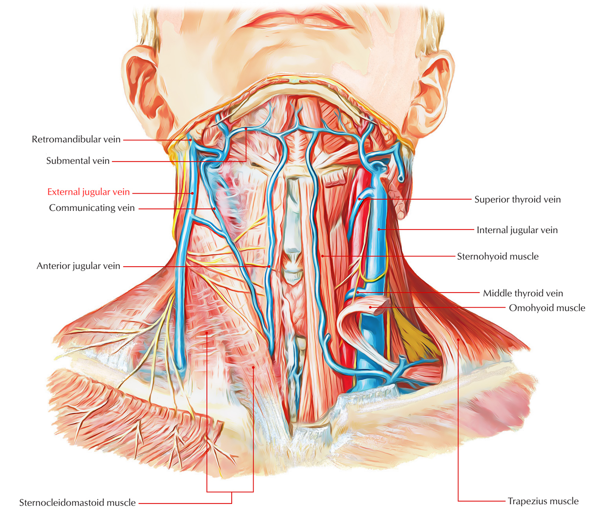
External Jugular Vein is among the main veins of the neck region. It is among the most superficial structures traveling from the posterior triangle of the neck.

External Jugular Vein
Origin
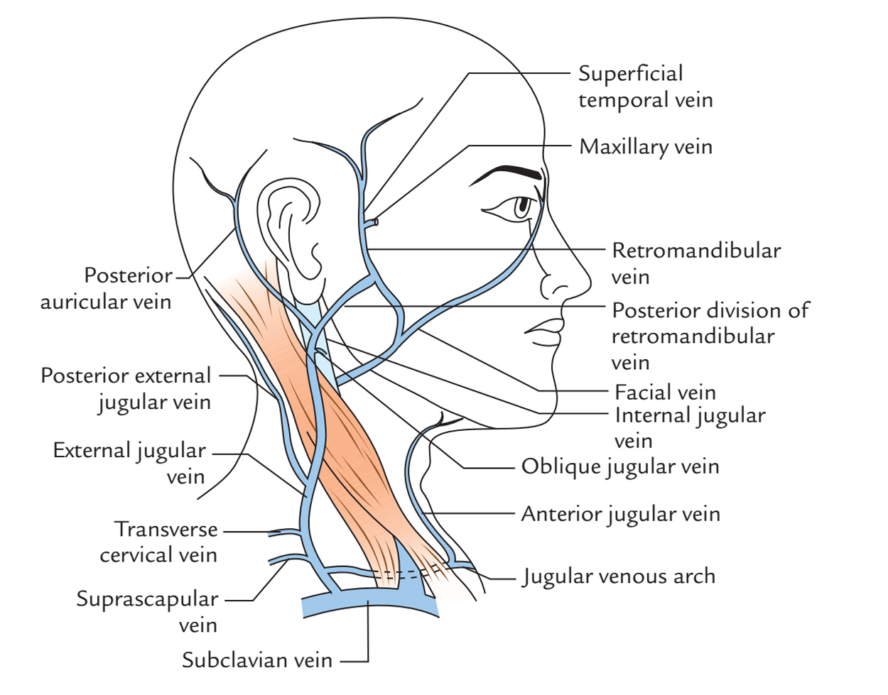
It originates just inferior to the angle of the mandible by the combination of posterior branch of retromandibular vein along with posterior auricular vein.
Insertion
It then travels nearly vertically downward through the sternocleidomastoid beneath the concealment of platysma in order to perforate the deep cervical fascia within the anteroinferior angle of the posterior triangle approximately 2.5 cm superior to the clavicle alongside the posterior margin of the sternocleidomastoid and move in the supraclavicular space. After it travels via this space it terminates inside the subclavian vein.
Structure
Valves
It has two pair of valves:
- The lower pair being located at its entrance into the subclavian vein.
- The upper in mostly about 4 cm above the clavicle.
The portion of vein between the two sets of valves is often dilated, and is termed the sinus. These valves do not prevent the regurgitation of the blood or the passage of injection from below upward.
Variation
The external jugular vein has significant number of variations in its size as well as course. It particularly becomes visible in old age as it impedes the venous return towards the right side of the heart and distends the vein, whenever one holds his breath or inflates his cheek with mouth closed.
Tributaries

External Jugular Vein: Tributaries
Formative
- Posterior auricular vein.
- Retromandibular vein.
Other
- Posterior external jugular vein.
- Oblique jugular vein.
Terminal
- Transverse cervical vein.
- Suprascapular vein.
- Anterior jugular vein.
Clinical Significance
Visibility of the External Jugular Vein
In children as well as women the external jugular vein is less visible, for the reason that compared to the tissue of men their subcutaneous tissue is thicker.
The vein may be hard to recognize in obese persons, even when they hold their breath, which blocks the venous return towards the right side of the heart and inflates the vein.
Due to long periods of raised intrathoracic pressure, the superficial veins of the neck are enlarged and every so often tortuous in professional singers.
As a Venous Manometer
Generally, when the patient is reclined at a horizontal angle of 30°, the external jugular vein act as venous manometer.
In the external jugular veins, the level of the blood extends approximately one third of the way upwards the neck.
The blood level falls as the patient sits up, until it is no longer visible behind the clavicle.
Catheterization
The external jugular vein can be used for catheterization; however valves and tortuosity may make the passage of the catheter challenging. It is the one most commonly used for the reason that the right external jugular vein is in the maximum direct alignment with the superior vena cava.
The catheterization of vein is done about halfway in the middle of the plane of the cricoid cartilage as well as the clavicle. The insertion of the catheter must be done during inspiration whenever the valves are open.

 (55 votes, average: 4.85 out of 5)