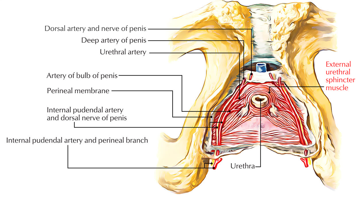
The external urethral sphincter emerges on the ischiopubic ramus and also via the opposite side enters within the intermeshing muscle fibers. It is controlled through the deep perineal section of the pudendal nerve, where action within the nerve fibers contracts the urethra.

External Urethral Sphincter
Structure
When around 500 ml of the urinary bladder volume is achieved, the pressure at this volume makes the internal sphincter to open up. Urine will go into the urethra now. Another sphincter is located around 2 cm distal within the urethra. This is referred to as the external urethral sphincter. This sphincter is comprised of skeletal muscle and as a result is subject to voluntary control. Till the suitable time comes, the control of this sphincter keeps the urine from leaving the urethra. The urinary bladder goes on to fill up and expand if it’s not emptied yet. The impulse to empty keeps getting stronger. Whenever the urinary bladder fills up to around 800 ml, now even for the external urethral sphincter the pressure is too immense. Regardless if it is the suitable time or not this sphincter is going to open now.
Storage Reflex
Storage is enhanced through bladder afferent activation of the Onuf’s nucleus, which improves the tone of the external urethral sphincter through the somatic pudendal nerve. The pontine storage center, within the lateral pons, similarly activates the Onuf’s nucleus along with the pudendal nerve output towards the external urethral sphincter. At the same time, acquiring input through pelvic afferents, the hypogastric nerve (sympathetic), when activating the internal urethral sphincter (bladder neck) suppresses the detrusor muscle and bladder ganglia, which has strong interpretation of alpha-1 receptors.
Emptying Reflex
Parasympathetic discharge via the pelvic nerve creates detrusor constriction and suppression of the internal sphincter (bladder neck), processes which help in emptying. The pontine micturition centre, which activates parasympathetic motor or “efferent” fibers whose cell bodies reside in the somatic pudendal nerve moderates the emptying. Furthermore, impulses from the pontine micturition centre also prevent sympathetic output from the hypogastric nerve, causing relaxation of the bladder neck, an activity that also assists in emptying. Also, signals via the pontine micturition centre also restrict the motor output towards the pudendal nerve, resulting in relaxation of the striated urethral sphincter, furthermore helping with emptying.
Variations in Sexes
Women compared to males, have a more elaborate external sphincter muscle as it is comprised of three components: the sphincter urethrae, urethrovaginal muscle, along with the compressor urethrae. The urethrovaginal muscle fibers envelop all around the vagina and urethra and constriction causes contraction of both of the vagina as well as the urethra. The beginning of the compressor urethrae muscle is the right and left inferior pubic ramus and also it encloses anteriorly over the urethra so whenever it constricts, it compresses the urethra in opposition to the vagina. The external urethrae like in males, covers around the urethra. Congenital defects of the female urethra may be surgically restored by vaginoplasty. Anteriorly, a group of muscle fibers cover the urethra and together create the external urethral sphincter. It covers the membranous portion of the urethra within male, and the urethra and vagina within female. Its superficial fibers emerge via the transverse perineal ligament and move backwards on each side of the urethra in order to be attached within the perineal body. Its deep fibers emerge through the inner sides of the ischiopubic rami and travel horizontally in order to encircle the urethra. The sphincter urethrae muscle creates the voluntary external urethral sphincter.
Functions
In males as well as females, both internal together with external urethral sphincters serve to suppress the release of urine. In males, the internal sphincter muscle of urethra functions to reduce reflux of spermatic fluids within the male bladder at the time of ejaculation.
Nerve Supply
Deep branch of Perineal Nerve.

 (47 votes, average: 4.51 out of 5)