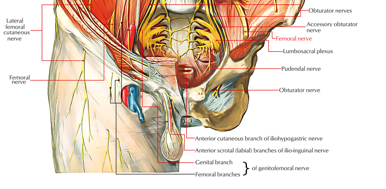
Femoral Nerve is the main nerve of anterior compartment of thigh. It originates from the dorsal sections of the anterior primary rami of L2, L3, L4 nerves and is the largest branch of lumbar plexus. It goes into the thigh behind the inguinal ligament and lateral to femoral sheath while descending via psoas major and appearing on its lateral order between psoas and illiacus.

Femoral Nerve
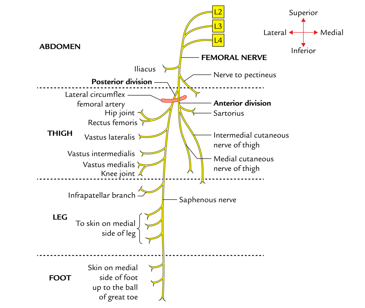
It digs into posterior and anterior section in femoral triangle 2 cm distal to the inguinal ligament. The lateral circumflex femoral artery is straddled by both sections. The illiacus in the abdomen and all the muscles of anterior compartment of the thigh is supplied by motor branches of it. The large cutaneous area on the anterior and medial part of thigh, medial side of leg and foot is supplied by the cutaneous branches of femoral artery. Hip and knee joints are also supplied by its articular branches.
Anterior section
It produces 2 cutaneous branches and 1 muscular branch.
(a) The cutaneous nerves are:
- medial cutaneous nerve of the thigh and
- intermediate cutaneous nerve of the thigh.
(b) The muscular branch provides the sartorius.
Posterior section
It produces 1 cutaneous branch, the saphenous nerve, and 4 muscular branches to provide the quadriceps femoris.
Course
It enters the femoral triangle by passing behind the inguinal ligament just lateral to the femoral artery. In the thigh, it lies in the groove between the iliacus and the psoas major, outside the femoral sheath, and lateral to the femoral artery. After a short course of about 2 cm below the inguinal ligament, the nerve divides into anterior and posterior divisions which are separated by the lateral circumflex femoral artery.
Branches and Distribution

Femoral Nerve: Branches
Muscular
(1) The anterior division supplies the sartorius; and
(2) the posterior division supplies the rectus femoris, the three vasti and the articularis genu. The articularis genu is supplied by a branch from the nerve to vastus intermedius.
Cutaneous
(1) The anterior division gives two cutaneous branches, the intermediate and the medial cutaneous nerves of the thigh: and
(2) The posterior division gives only one cutaneous branch, the saphenous nerve. These nerves have been described earlier.
Articular
(1) The hip joint is supplied by the nerve to the rectus femoris; and
(2) the knee joint is supplied by the nerves to the three vasti. The nerve to the vastus medialis contains numerous proprioceptive fibres from the knee joint, accounting for the thickness of the nerve. This is in accordance with Hilton’s law: Nerve supply to a muscle which lies across a joint, not only supplies the muscle, but also supplies the joint beneath and the skin overlying the muscle.
Vascular
To the femoral artery and its branches.
Note: The nerve to the pectineus arises from the medial side of the femoral nerve Just above the inguinal ligament. It passes obliquely downwards and medially, behind the femoral sheath, to reach the anterior surface of the muscle.
Clinical Significance
Injury of The Femoral Nerve
It’s uncommon but may be injured by a stab, gunshot wounds, or a pelvic fracture. Listed here are the characteristic clinical features:
Motor loss.
- Poor flexion of the thigh, because of paralysis of the iliacus and sartorius muscles.
- Inability to extend the knee, because of paralysis of the quadriceps femoris.
Sensory decrease
- Sensory decline over the anterior and medial aspects of the thigh, as a result of engagement of the intermediate and lateral cutaneous nerves of the thigh.
- Sensory loss on the medial side of the leg and foot up to the ball of the great toe (first metatarsophalangeal joint), because of engagement of the saphenous nerve.
Femoral Nerve Neuropathy
The primary trunk of the femoral nerve isn’t subject to an entrapment neuropathy . However, it might be compressed by the retroperitoneal tumors. A localized neuropathy of the femoral nerve may happen in diabetes mellitus. Listed here are the characteristic clinical features:
- Wasting and weakness of quadriceps resulting in significant trouble in walking.
- Pain and paraesthesia on the anterior and medial aspects of the thigh going down along the medial aspect of the leg and foot along the distribution of the saphenous nerve.

 (51 votes, average: 4.69 out of 5)