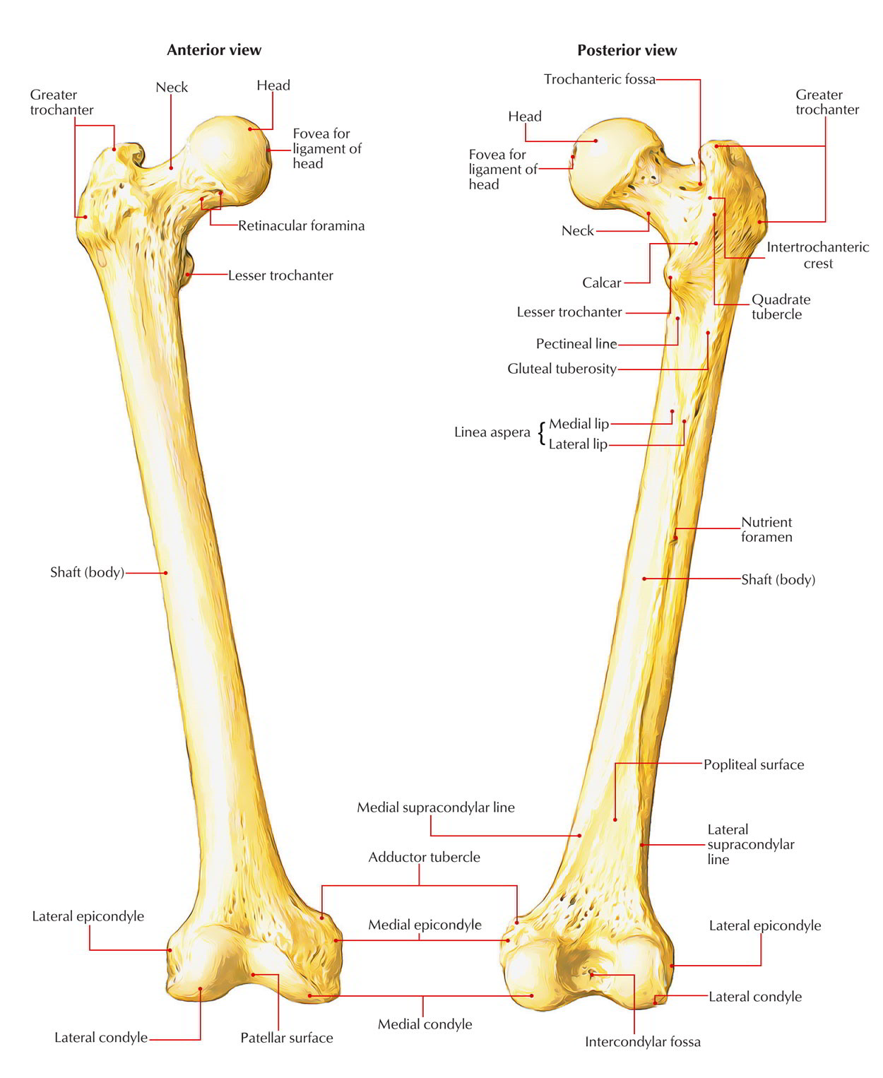
The Femur is the longest and strongest bone of the body, present in the thigh (Latin femur = thigh). It’s around 18 inches (45 long), i.e., about quarter of the height of the individual. At the upper end it articulates with the hip bone to create the hip joint, and at the lower end it articulates with the patella and tibia. The femur conducts body weight from the hip bone to the tibia in standing position.
Femur
Parts
- The femur is the long bone and is composed of 3 parts: upper end, shaft, and lower end.
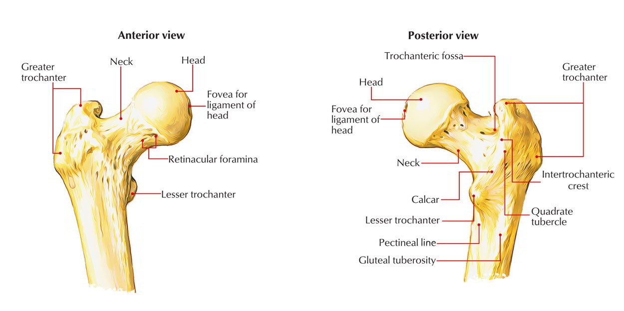
- The upper end contains the head, neck, and lesser and greater trochanter.
- The shaft of the femur is gradually convex anteriorly with maximum convexity in the middle third where the shaft is narrowest.
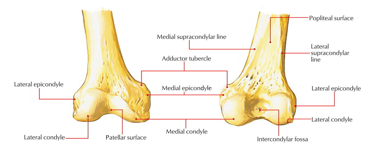
- The lower end of the femur is enlarged to create medial and lateral condyles. Both condyles project backwards and are divided by the intercondylar fossa. The most notable points on the condyles are named epicondyles.
- The shape of femur departs markedly from other long bones in the meaning the long axis of head and neck projects superomedially at an angle to the long axis of the shaft of bone. This angle enables greater freedom in the hip joint but inflicts great strain on the neck during transmission of weight, which makes it susceptible to fracture.
Side Determination
- The upper end bears a rounded head whereas the lower end is widely expanded to form two large condyles.
- The head is directed medially.
- The cylindrical shaft is convex forwards.
Anatomical Position
- The head is directed medially upwards and slightly forwards.
- The shaft is directed obliquely downwards and medially so that the lower surfaces of the two condyles of the femur lie in the same horizontal plane.
Points to be Noticed
- Angle of inclination of femur: It’s an angle between the long axes of neck and shaft of the femur thus also named neck-shaft angle. The normal neck-shaft angle is 125 ° in adults and 160 ° in youngsters. It’s less in female.
- Angle of femoral torsion: It’s an angle between long axis of the head and neck of femur, and transverse axis of the femoral condyles. It measures about 7 ° in men and 12 ° in female).
Upper End

Femur: Head
Head
- It creates about two-third of a sphere and articulates with the acetabulum of the hip bone to create the hip joint.
- It presents a small pit, the fovea, just below and behind the center, which gives connection to the ligament of the head of femur (ligamentum teres femoris).
Neck
- It’s 5 cm long and attaches the head with all the shaft.
- It’s directed upward, medially, and somewhat forwards.
- The angle between its lower border and the medial border of shaft is termed neck-shaft angle.
- The neck presents 2 edges- upper and lower, and 2 surfaces- anterior and posterior.
Upper Border
- It’s concave and horizontal.
- It meets the shaft near the greater trochanter.
Lower Border
- It’s straight and oblique.
- It meets the shaft near the lesser trochanter.
Anterior Surface
- It’s flat and bears a number of oblique bony ridges.
- It meets the shaft at intertrochanteric line.
- It’s entirely intracapsular.
Intertrochanteric Line
- It continues downward and medially below the lesser trochanter on the posterior aspect of femur as spiral line.
- It gives connection to 2 ligaments and 2 muscles:
- Capsule of the hip joint.
- Iliofemoral ligament (strongest ligament in the body).
- Vastus lateralis to its upper end.
- Vastus medialis to its lower end.
Posterior Surface
- It’s convex from above downward and concave from side to side.
- It meets the shaft at the intertrochanteric crest.
- Its medial half is intracapsular.
- It presents a dim groove, which lodges the tendon of obturator externus.
Intertrochanteric Crest
- It goes from the posterosuperior angle of greater trochanter to the tip of lesser trochanter.
- It presents a rounded tubercle near the middle named quadrate tubercle, which gives insertion to the quadratus femoris.
The neck attaches the head to the shaft about 5 cm away from the hip joint. This allows the lower limb to swing clear of the pelvis. Therefore, the neck of femur is comparable to the clavicle in the upper limb.
Greater Trochanter
- It’s a quadrilateral elevation, projecting upward from the lateral aspect of the junction of neck and shaft.
- It presents 1 border (upper) and 3 surfaces (anterior, medial, and lateral).
Upper Border
Its posterior part presents the apex or tip of greater trochanter, which gives connection to the piriformis.
Anterior Surface
- It gives connection to the gluteus minimus on a ridge in its lateral part. Its medial part is related to a synovial bursa- trochanteric bursa of gluteus minimus.
Medial Surface
It presents 2 melancholy:
- A depression (trochanteric fossa) at its junction together with the neck for insertion of obturator externus.
- A shallow depression above and in front of trochanteric fossa for insertion of obturator internus together with the gemellus superior and gemellus inferior.
Lateral Surface
It’s quadrilateral and split diagonally by an oblique ridge into the upper and lower triangular regions.
- The ridge gives connection to the gluteus medius muscle.
- The triangular regions- anterior and posterior to the ridge are associated with the trochanteric bursae of the gluteus medius and gluteus maximus, respectively.
Lesser Trochanter
- It’s a conical projection rising from the posteromedial surface of the neck-shaft angle. It’s directed medially.
- Its apex gives connection to the psoas major.
- Iliacus is connected to its base on the front.
The connection of fibrous capsule at the upper end of femur: Anteriorly it’s connected to the intertrochanteric line but posteriorly it’s connected about 1 cm medial to the intertrochanteric crest. Thus, the lateral part of the posterior outermost layer of the neck of femur is extracapsular and its medial half is intracapsular.
Shaft

Femur: Shaft
Features
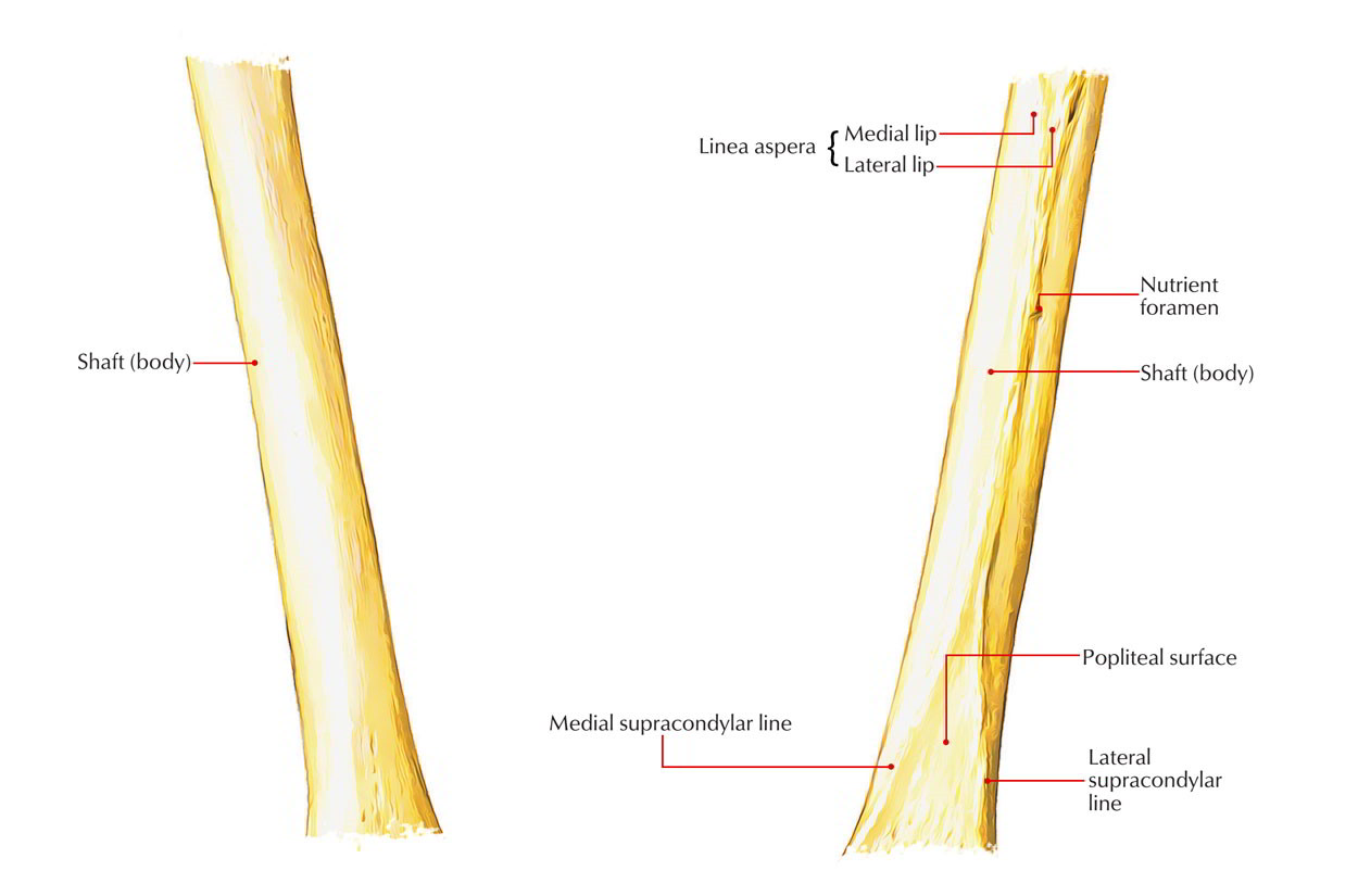
- Essentially the shaft of femur presents 3 surfaces – anterior, medial, and lateral and 3 edges – medial, lateral, and posterior.
- The middle third of posterior border is represented by a thick ridge termed linea aspera, which functions as a buttress to withstand the compressive forces and prevents the anterior bowing of the shaft. The linea aspera presents inner and outer lips and an interceding intermediate area. When the upper end of linea aspera is tracked upward, its inner and outer lips diverge and enclose a triangular posterior surface in the upper one-third of the shaft. The inner (medial) lip becomes constant with the coil line and the outer (lateral) lip becomes constant with the gluteal tuberosity, which goes up to the root of greater trochanter.
- When the lower end of linea aspera is followed downward, its inner and outer lips, diverge below, and enclose a triangular popliteal surface in the lower one-third of the shaft. The inner (medial) lip becomes constant with the medial supracondylar line, which finishes at the adductor tubercle. The outer (lateral) lip becomes constant with Lateral supra condylar line. So, strictly speaking, the shaft presents 5 surfaces- anterior, medial, lateral, upper posterior, and lower posterior (popliteal surface).
Borders and Surfaces of the Shaft of Femur
| Part of femur | Borders | Surfaces |
|---|---|---|
| Middle 1/3rd of the shaft | Medial border | Anterior surface |
| Lateral border | Medial surface | |
| Posterior border (linea aspera) | Lateral surface | |
| Upper 1/3rd of the shaft | Medial border | Anterior surface |
| Lateral border | Medial surface | |
| Spiral line | Lateral surface | |
| Gluteal tuberosity | Posterior surface | |
| Lower 1/3rd of the shaft | Medial border | Anterior surface |
| Lateral border | Medial surface | |
| Medial supracondylar line | Lateral surface | |
| Lateral supracondylar line | Popliteal surface |
Attachments
The shaft of femur gives connection to these muscles and intermuscular septa:
- Attachments of intermuscular septa: 1. Medial intermuscular septum is connected to the medial lip of linea aspera. 2. Lateral intermuscular septum is connected to the lateral lip of the linea aspera.
- Muscular attachments:
- Vastus intermedius appears from the upper three fourth of anterior surface and adjoining lateral surface.
- Articularis genu originates by few small slides from the lower quarter of anterior surface immediately below the vastus intermedius.
- Vastus lateralis appears in a linear manner from the upper part of intertrochanteric line, anterior and inferior edges of greater trochanter, lateral margin of gluteal tuberosity, and lateral lip of linea aspera.
- Vastus medialis appears in a linear manner from the lower part of intertrochanteric line, coil line, and the medial lip of linea aspera and the upper 1 fourth of medial supracondylar line.
- Gluteus maximus is added into the gluteal tuberosity.
- Adductor longus is added into the medial lip of linea aspera.
- Pectineus is added into a line going from the lesser trochanter to the upper end of linea aspera.
- Adductor brevis is added into a line going from the lesser trochanter to the upper part of linea aspera.
- Adductor magnus is added into the medial margin of gluteal tuberosity, linea aspera, medial supracondylar line, and adductor tubercle.
- Gastrocnemius: The medial head originates from the popliteal surface just above the medial condyle. The lateral head originates primarily from the lateral condyle but also stretches over the lower end of lateral supracondylar line.
- Plantaris rises from the lower end of lateral supracondylar line just above the lateral head of gastrocnemius.
- The arrangement of 9 structures connected to the lineaaspera: From medial to lateral side, these are:
- Vastus medialis.
- Medial intermuscular septum.
- Adductor brevis (in the upper) and adductor longus (in the lower part).
- Adductor magnus.
- Short head of biceps femoris.
- Posterior intermuscular septum.
- Vastus lateralis.
- Vastus intermedius.
Lower End

Femur: Lower End
Medial Condyle
- Its most notable point is named medial epicondyle, which gives connection to the upper end of medial collateral ligament.
- A projection posterosuperior to the medial epicondyle is known as adductor tubercle, which gives insertion to the ischial head of adductor magnus.
- Its lateral surface creates medial boundary of intercondylar fossa.
Lateral Condyle
- It’s stouter and more powerful in relation to the medial condyle but less notable.
- Its lateral surface presents a bulge referred to as lateral epicondyle which gives connection to the fibular collateral ligament of the knee joint.
- Smooth opinion above and behind the lateral epicondyle there is an origin to the lateral head of gastrocnemius.
- A groove below and behind the lateral epicondyle gives connection to popliteus in its anterior part. Tendon of popliteus takes up the posterior part of the groove during total flexion in the knee joint.
- Its medial surface creates the lateral boundary of intercondylar fossa.
Intercondylar Fossa (Intercondylar Notch)
- It’s a deep notch, which divides 2 condyles posteriorly.
- It’s restricted posteriorly above by intercondylar path.
- It presents medial and lateral walls and floor.
- Medial wall of the fossa gives attachments to the upper end of posterior cruciate ligament in its anteroinferior part.
- Lateral wall of the fossa gives connection to the upper end of anterior cruciate ligament in its posterosuperior part.
Mnemonic: LAMP = Lateral condyle gives connection to Anterior cruciate ligament and Medial condyle to Posteriorcruciate ligament.
Articular Surface
The lower end of femur presents a V-shaped articular surface inhabiting the anterior, inferior, and posterior surfaces of both condyles.
- The apex of ‘V’ is referred to as patellar surface which inhabits the anterior surfaces of 2 condyles and articulates with the patella.
- The 2 connects of ’V’ create tibial surfaces which inhabit the inferior and posterior surfaces of 2 condyles to come in contact with corresponding articular surfaces of tibial condyles.
- The patellar surface is saddle-shaped. Its lateral portion is wider and extends to a higher level in relation to the medial portion, corresponding to articular outermost layer of the patella.
Connection of fibrous capsule at the lower end of femur:
- Posteriorly, it’s connected to the intercondylar line and articular margins.
- Anteriorly, it’s deficient for communication of suprapatellar bursa together with the synovial cavity of knee joint.
- Laterally and medially, it’s connected along a line 1 cm above the articular margins. It’s essential to note the groove for popliteus is intracapsular.
Clinical Significance
Fracture Neck of Femur
It’s quite common in aged especially in women because of osteoporotic changes in the neck.
Types of Fracture
The fracture might be intracapsular (subcapital, transverse cervical) or extracapsular (basal, intertrochanteric, and subtrochanteric). In intracapsular fracture, the retinacular boats- the main source of blood supply to the head- are injured. This results in delayed healing or nonunion of fracture, or even avascular necrosis of the head of femur. In intracapsular fracture of the neck of femur, the affected limb is shortened and characteristically held in laterally rotated position with the toes pointing laterally.
Ossification
The femur ossifies from 5 centers: 1 primary and 4 secondary centers. The primary center appears in the midshaft. 3 secondary centers show up in the upper end and 1 secondary center in the lower end. The age of look and fusion of these centers is provided in Table below. Ossification of the femur.
| Centres | Time of appearance | Time of fusion |
|---|---|---|
| Primary centre | 7th to 8th week of IUL | |
| In mid shaft | ||
| Secondary centres | ||
| 1 for head | 1 year | Three separate epiphysis 18th year |
| 1 for greater trochanter | 3 years | |
| 1 for lesser trochanter | 13 years | |
| For lower end 1 | 9 months (at the time of birth) | 20 years |
Clinical Significance
Medicolegal importance of ossification center at the lower end of femur: The secondary center at the lower end of femur is exceptional in the meaning it seems during beginning/ just before arrival (ninth month of IUL). It’s of medicolegal significance because its look in radiograph signifies maturity of the fetus.
Test Your Knowledge
Femur Quiz


 (53 votes, average: 4.64 out of 5)