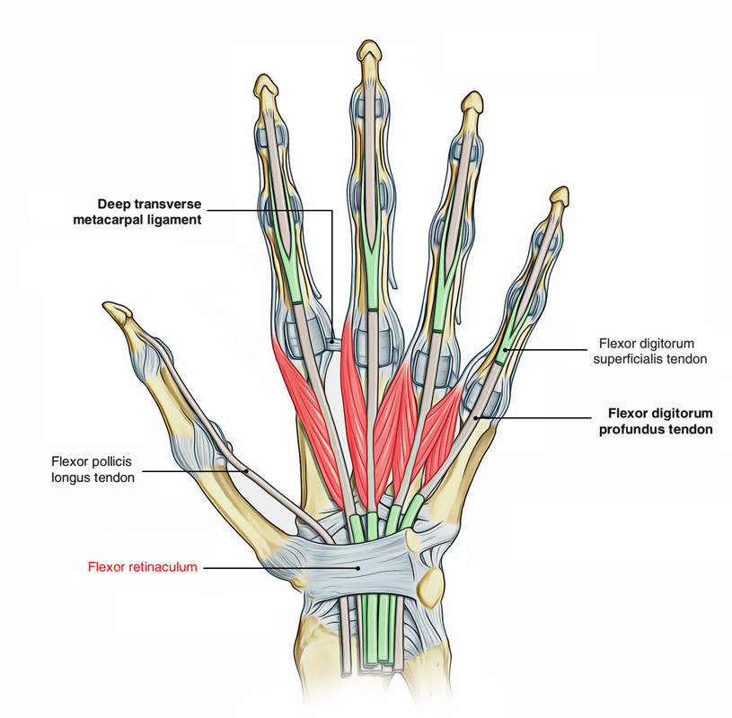
It is rectangular in shape and has roughly the shapes and size of a postage stamp.Like a postage stamp, it presents two surfaces and four borders.The flexor retinaculum is a robust fibrous band created by the thickening of deep fascia in front of the carpus (anatomical wrist).
Attachment
- The flexor retinaculum of the hand attaches to the hamate bone’s hamulus, which is a curved process that lies on the bottom of the hamate bone.
- It connects to the middle of the pisiform, which is a small wrist bone that is shaped like a pea.
- In addition, it connects laterally to the scaphoid and across the middle of the trapezium.
- With carpus, it forms an osseofibrous tunnel called carpal tunnel for the passage of flexor tendons of the digits.

Flexor Retinaculum
Stucture
- The flexor retinaculum consists of three parts. By its attachment to the pisiform, hamate hook, scaphoid tuberosity, and ridge of trapezium, the central part is specified.
- The hamate hook lies at the apex of this interval. An interval exists in between these fibers and those that emerge from the hypothenar muscles. This portion of the flexor retinaculum is 1.24 ± 1.3 cm in length.
- The fibers arise from the pisiform to form a distinct head, on the ulnar element the majority of the radial fibers originate from the trapezium deep to the thenar musculature. A narrow strip of fibers emerges from the scaphoid tubercle.
- The proximal section of the flexor retinaculum has irregular boundaries. This section is merged to and indivisible from the ante-brachial fascia. The density of this section varies considerably amongst various specimens.
- The distal section of the flexor retinaculum consists of an aponeurosis between the thenar and hypo-thenar muscles. The thenar muscles (flexor pollicis brevis muscle and to a lesser degree abductor pollicis brevis and opponens pollicis) originate from the radial side of this aponeurosis while, the hypothenar muscles (oppo- nens digiti minimi and flexor digiti minimi brevis) originate from the ulnar element of the aponeurosis. This section of the flexor retinaculum has a mean length of 0.99 ± 0.13 cm.
Clinical Significance
Carpal Tunnel Syndrome
If the flexor retinaculum compresses the median nerve, carpal tunnel syndrome may occur. Symptoms consist of:
- Tingling.
- numbness, and.
- pain in the wrists, hands, and forearm.
Carpel tunnel syndrome might be caused by anything that causes inflammation in the wrist.
In severe cases, treatment requires surgery to divide the flexor retinaculum. Sometimes, it may be connected to other conditions, such as arthritis, or repetitive actions, like typing.
Risk Factors
- Kinds of work that are related include computer work, deal with vibrating tools, and work that requires strong grip.
- Risk factors consist of obesity, recurring wrist work, pregnancy, and rheumatoid arthritis.
- There is tentative proof that hypothyroidism increases the danger. It is unclear if diabetes plays a role.
- Diagnosis is suspected based upon indications, symptoms, and particular physical tests and may be validated with electrodiagnostic tests.
- If muscle losing at the base of the thumb exists, the diagnosis is likely.
Treatment
- Physically activities can decrease the threat of establishing CTS.
- Symptoms can be improved by using a wrist splint or with corticosteroid injections.
- Surgery to cut the transverse carpal ligament works with better results at a year compared to non-surgical choices.
- Further splinting after surgery is not needed.
x

 (54 votes, average: 4.66 out of 5)