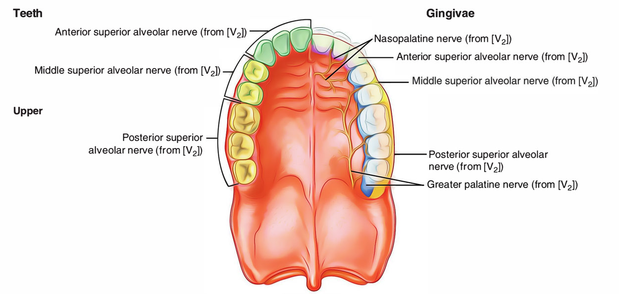
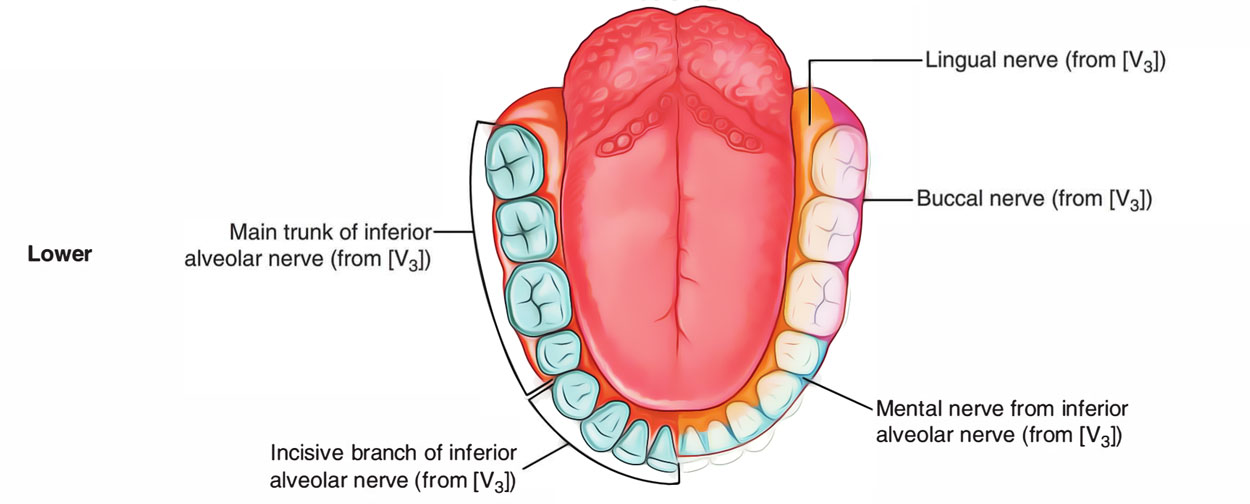
The gums are made up of fibrous tissue covered with a smooth vascular mucous membrane. They cover the alveolar protuberances of the jaws and the necks of the teeth. The fibrous tissue of gum is constant with the gum membrane which connects the teeth to their plugs at the stems of the teeth.

Gingiva
Parts of Gum
The gum/gingiva has three parts.
- Free part a.k.a. free gingiva, which envelops the neck of tooth like a collar.
- Affixed part a.k.a. attached gingiva, which is strongly connected to the alveolar procedure.
- Interdental portion a.k.a. interdental gingiva, which is the expansion of the connected gingiva in the middle of the teeth.
Structure
Marginal (Free) Gingiva
- This part of the gingiva envelops the neck of the tooth and is not directly connected to the tooth and creating the soft tissue wall of the gingival sulcus.
- It expands via the gingival margin towards the gingival groove.
- The free gingival margin itself is located 1.5 to 2 mm coronal or towards the head to the cementoenamel junction following tooth eruption and is about 1 mm thick.
Gingival Groove
- A shallow line or indentation is found on the surface of the gingiva that spllits the free gingiva via the attached gingiva is present around 50% of the time.
- The gingival groove commonly shows the location of the base of the gingival sulcus.
Keratinized Gingiva
- The band of keratinized gingiva expands via the gingival margin to the mucogingival junction.
- An increased vulnerability to tissue breakdown might exist when pulling of the lip or cheek lead to movement of the free gingival margin or papilla that is measured by the favorable pull test.
- An appropriate width of connected gingiva might then be specified as the quantity of keratinized tissue is required to aid the conserve a non-inflamed steady gingival margin.
- The apicocoronal width of the keratinized gingiva differs via less than 1 to 9 mm and is usually largest in the anterior maxilla and posterior lingual mandible.
- A narrow zone of keratinized gingiva is often also found in specific teeth. These teeth consist of mandibular canines and premolars, prominent teeth, and teeth connected with unusual frenum or muscle connections.
Connected Gingiva
- It is the section of the gingiva that extends to the top via the area of the free gingival groove towards the mucogingival junction.
- In the absence of inflammation the facial connected gingiva is specified plainly.
- In the hard palate, there is no clinical separation in the middle of the connected gingiva and the residing masticatory mucosa.
- Connected gingiva is usually covered by keratinized or parakeratinized epithelium with significant ridges.
- This tissue is developed in order to endure the rigors of chewing, tooth brushing, and oilier operational tensions, and it is securely bound towards the main tooth and bone.
Nerve Supply

Gingiva Innervation
Upper gums
- The labial part is supplied by posterior, middle, and anterior-superior alveolar nerves.
- The lingual part is supplied by greater palatine and nasopalatine nerves.
Lower gums
- The labial part is supplied by buccal branch of mandibular nerve, and incisive branch of mental nerve.
- The lingual part is supplied by lingual nerves.
Lymphatic Drainage
- Upper gums: The lymphatics via upper gums drain into submandibular lymph nodes.
- Lower gums: The lymphatics via anterior part a.k.a. gums of lower central incisors drain into submental lymph nodes while those via remaining part drain into submandibular lymph nodes.
Clinical Significance
- Gingivitis: inflammation of gums or gingivitis and suppuration with formation of pockets of pus in between teeth and gums causing clinical condition called pyorrhea, might be triggered by the inappropriate oral health which clinically present as:
- o Discharge of pus at the margins of gums.
- o Foul odor throughout breathing.
- Scurvy: the gums become inflamed, spongy, and bleed on touch in scurvy because of shortage of vitamin C.
x

 (56 votes, average: 4.90 out of 5)