Gluteal Region is the back and side of lateral half of pelvic region. It covers the area from iliac crest from above to the gluteal fold below. It goes laterally up to a virtual line converging the anterior superior iliac spine to the anterior edge of higher trochanter and Medially goes up to mid-dorsal line and natal cleft. It is the most common site of intra-muscular injections
Surface Landmarks
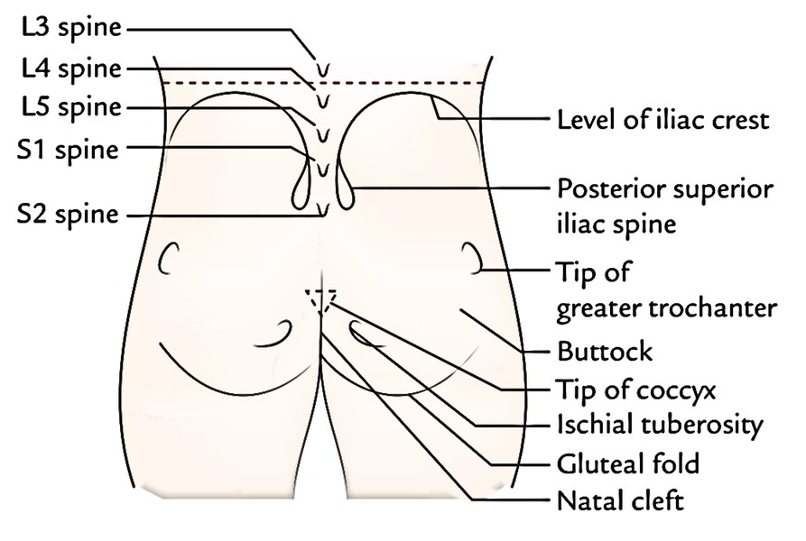
- Buttock: It’s a round bulge on the posterior aspect of the gluteal region.
- Gluteal fold: It’s a transverse skin crease, which creates the lower limit of the gluteal region. It’s generated by a linear adherence of the skin to the deep fascia, obliquely across the inferior border of gluteus maximus.
- Natal cleft: It’s a midline cleft between the 2 buttocks, which starts at the level of the third spine of the sacrum and deepens inferiorly with lower sacral spines and coccyx being located in its floor.
- Coccyx: It is located just behind the anal orifice and can be recognized by its comparative freedom under pressure.
- Posterior superior iliac spine (PSIS): It is located in a skin dimple in the level of second sacral (S2) spine.
- Ischial tuberosity: It’s a rounded bony mass on which 1 sits. It is located below the PSIS in precisely the same vertical plane at a lower level in relation to the tip of coccyx. It can be felt by pressing your fingers upward into the medial part of the gluteal fold. It’s 5 cm above the gluteal fold and about same distance from the midline.
- Tip of greater trochanter: It is located just in front of hollow on the side of hip, about 1 hand’s width below the tubercle of iliac crest.
- Iliac crest: It can be felt as a curved bony ridge in a groove at the lower limit/margin of the waistline. The maximum point of iliac crest corresponds to the interval between the spines of L3 and L4 vertebrae.
Key Points
- Nelaton’s line: It’s the line joining the anterior superior iliac spine and the most notable point of ischial tuberosity. It crosses the tip of greater trochanter.
- Bryant’s triangle: With the patient in supine position, first draw a vertical line passing downwards from the anterior superior iliac spine (ASIS) and now draw a line going from ASIS to the tip of greater trochanter (spinotrochanteric line). Lastly draw a horizontal line from the tip of greater trochanter to the very first line. The triangle so created is referred to as Bryant’s triangle.
Superficial Fascia
The superficial fascia in the gluteal region is thick and includes considerable subcutaneous fat especially in adult females, that is responsible for a characteristic round contour of the buttock in them.
Cutaneous Nerves
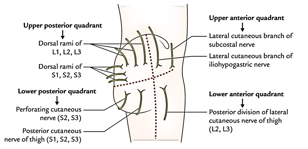
- The cutaneous nerves of the gluteal region are originated from several sources, and converge in this region from all the directions. The cutaneous innervation of the gluteal region is split into 4 quadrants- upper anterior, upper posterior, lower anterior, and lower posterior.
- Upper anterior quadrant is supplied by the:
1. lateral cutaneous branch of subcostal nerve (T12), and
2. lateral cutaneous branch of iliohypogastric nerve (L1). - Upper posterior quadrant is supplied by the:
1. cutaneous branches from dorsal rami of upper 3 lumbar nerves (L1, L2, L3) and upper 3 sacral nerves (S1, S2, S3). - Lower anterior quadrant is supplied by the:
1. posterior section of lateral cutaneous nerves of the thigh (L2, L3). - Lower posterior quadrant is supplied by the:
1. posterior cutaneous nerves of the thigh (S1, S2, S3), and
2. perforating cutaneous nerves (S2, S3).
Cutaneous Arteries And Lymph Vessels
The cutaneous arteries supplying the gluteal region are originated from the superior and inferior gluteal arteries.
The lymph vessels from the gluteal region drain into the lateral group of superficial inguinal lymph nodes.
Deep Fascia
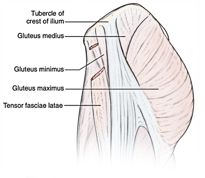
The deep fascia of the gluteal region is connected above to the iliac crest and behind to the sacrum. It carves twice along the iliac crest, first time to enclose the tensor fasciae latae and second time to enclose the gluteus maximus. Between tensor fasciae latae and gluteus maximus is a thick fascial sheet named gluteal aponeurosis which covers the gluteus medius.
Gluteal Ligaments
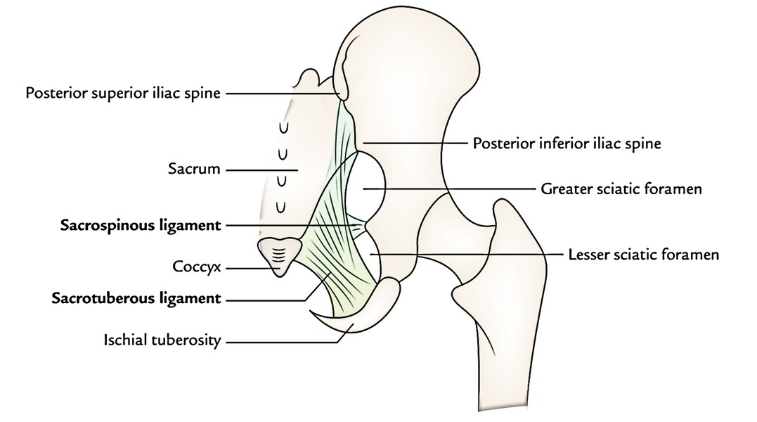
It’s of extreme significance for the people to study 2 significant gluteal ligaments (sacrotuberous and sacrospinous ligaments) in the gluteal region till they carry on to study the deep structures in this region.
Sacrotuberous Ligament
It’s broad band of fibrous tissue which stretches from sides of the sacrum and coccyx to the medial side of the ischial tuberosity.
Sacrospinous Ligament
It’s triangular sheet of fibrous tissue which stretches from ischial spine to side of the sacrum and coccyx.
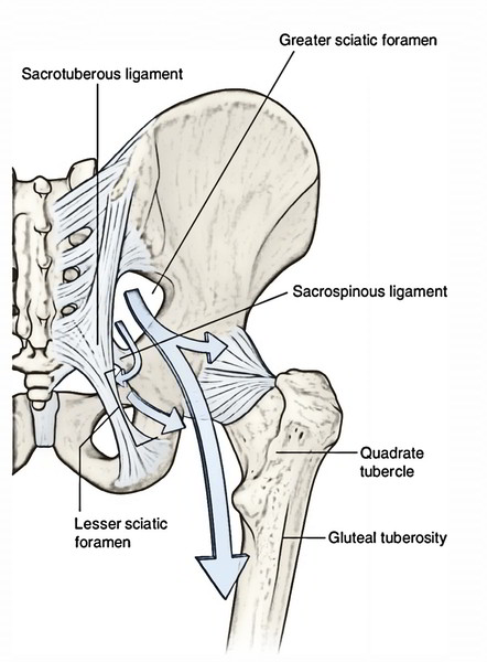
The sacrotuberous and sacrospinous ligaments convert the higher sciatic notch and lesser sciatic notch into greater sciatic foramen and lesser sciatic foramen, respectively.
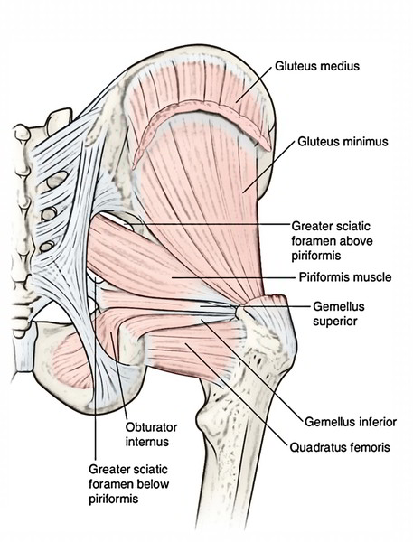
- The greater sciatic foramen is a passageway for structures leaving the pelvis and entering the gluteal region (example, sciatic nerve, superior and inferior gluteal vessels, etc.).
- The lesser sciatic foramen is a passageway for structures going into the perineum (example, pudendal nerve and artery).
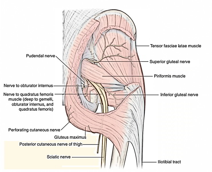
The greater sciatic foramen is regarded as the “door of the gluteal region” via which all arteries and nerves goes into the gluteal region from the pelvis.
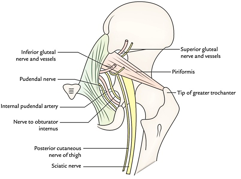
The structures going through the greater sciatic foramen are as follows:
- Piriformis: It appears from the pelvis and just about entirely fills the foramen. It’s the key muscle of the region.
- Structures passing below the piriformis:
1. Inferior gluteal nerve and vessels.
2. Sciatic nerve (most lateral structure).
3. Posterior cutaneous nerve of the thigh.
4. Nerve to quadratus femoris.
5. Pudendal nerve.
6. Internal pudendal vessels.
7. Nerve to obturator internus.
The past 3 structures cross the dorsal aspect of ischial spine and adjoining part of sacrospinous ligament (with internal pudendal artery between the nerves to obturator internus laterally and pudendal nerve medially), and arch forwards to goes into the perineum. The pudendal nerve and internal pudendal vessels run in the pudendal canal.
The structures going through the lesser sciatic foramen are as follows:
- Tendon of obturator internus.
- Nerve to obturator internus.
- Internal pudendal vessels.
- Pudendal nerve.
Muscles of The Gluteal Region
The muscles of the gluteal region are split into 2 groups- major and minor.
The major muscles of gluteal region are bigger in size and set superficially. They’re primarily extensor, abductors, and medial rotators of the thigh.
All these are 4 in number as given below:
- Gluteus maximus.
- Gluteus medius.
- Gluteus minimus.
- Tensor fasciae latae.
The minor muscles of gluteal region are smaller in size and set deeply under cover of the lower part of the gluteus maximus. They may be lateral rotators of the thigh and help stabilize the hip joint.
All these are 6 in number as follows:
- Piriformis.
- Superior and inferior gemelli.
- Obturator internus.
- Quadratus femoris.
- Obturator externus.
Attachments, Nerve Supply, and chief Actions of the Muscles of the gluteal region.
| Muscle | Origin | Insertion | Nerve supply | Actions |
|---|---|---|---|---|
| Gluteus maximus (quadrilateral muscle) | 1. Gluteal surface of the ilium behind posterior gluteal line 2. Outer slope of the dorsal segment of ilium 3. Dorsal surfaces of the sacrum and ilium 4. Sacrotuberous ligament | 1. 3/4th of the muscle into the iliotibial tract 2. 1/4th of the muscle into the gluteal tuberosity | Inferior gluteal nerve (L5; S1, S2) | 1. Chief extensor of the hip joint 2. Assists in getting up from sitting position |
| Gluteus medius (fan-shaped muscle) | Gluteal surface of the ilium between anterior and posterior gluteal lines | Oblique ridge on the lateral surface of the greater trochanter | Superior gluteal nerve (L5; S1) | 1. Abductor of the hip joint 2. Prevents the sagging of pelvis on the unsupported side |
| Gluteus minimus (fan-shaped muscle) | Gluteal surfaces of the ilium between anterior and inferior gluteal lines | Ridge on the lateral part of the anterior and inferior gluteal lines trochanter | ||
| Tensor fasciae latae (fusiform muscle) | Outer lip of the anterior part of iliac crest (from ASIS to tubercle) | Iliotibial tract | Superior gluteal nerve | Supports the femur on tibia during standing position |
| Piriformis | Piriformis | Piriformis | Piriformis | Piriformis |
| (pear-shaped muscle, Latin pirum = pear) | Pelvic surface of the middle three pieces of sacrum by three digitations | Apex/tip of greater trochanter | Ventral rami of S1, S2 | Lateral rotator of the thigh at hip joint |
| Gemellus superior | Posterior surface of the ischial spine | Medial surface of greater trochanter along with tendon of obturator internus | Nerve to obturator internus (L5; S1, S2) | Lateral rotator of the thigh at hip joint |
| Gemellus inferior | 1. Upper part of the ischial tuberosity 2. Lower part of greater sciatic notch | Same as that of gemellus superior | Nerve to quadratus femoris (L4; L5, S1) | Lateral rotator of the thigh at hip joint |
| Obturator internus (fan-shaped muscle) | Pelvic surface of the obturator membrane and surrounding bones | Medial surface of greater trochanter of femur in front of trochanteric fossa | Nerve to obturator internus (L5; S1) | Lateral rotator of the thigh at hip joint |
| Quadratus femoris (quadrilateral muscle) | Lateral border of the ischial tuberosity | Quadrate tubercle on the intertrochanteric crest and area below it | Nerve to quadratus femoris (L5; S1) | Lateral rotator of the thigh at hip joint |
Arteries of The Gluteal Region
The arteries of the gluteal region are as follows:
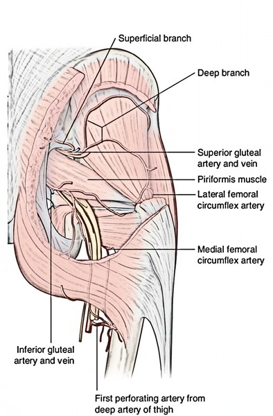
- Superior gluteal artery.
- Inferior gluteal artery.
- Internal pudendal artery.
Superior Gluteal Artery
The superior gluteal artery is a branch of the posterior section of internal iliac artery. It enters the gluteal region via greater sciatic foramen above the piriformis together with the superior gluteal nerve. Here it breaks up into the superficial and deep branches. The superficial branch enters between gluteus medius and maximus, and supplies both of them. The deep branch enters laterally between gluteus medius and minimus, and subdivides into upper and lower branches. The upper branch takes part in the formation of spinous anastomosis near the anterior superior iliac spine and the lower branch takes part in the formation of trochanteric anastomosis.
Inferior Gluteal Artery
The inferior gluteal artery is a branch of anterior section of the internal iliac artery and enters in the gluteal region below the piriformis together with the inferior gluteal nerve and continues together with the posterior cutaneous nerve of the thigh. It produces 3 sets of branches:
- Muscular branches to the adjacent muscles.
- Anastomotic branches to cruciate and trochanteric anastomoses.
- Artery to sciatic nerve (Latin arteria nervi ischiadici) accompanies the sciatic nerve and sinks into its substance to supply it. It’s the remnant of the axis artery of the lower limb.
Nerves of The Gluteal Region
The nerves of the gluteal region are as follows:
- Superior gluteal nerve.
- Inferior gluteal nerve.
The other nerves in the gluteal region are:
- Sciatic nerve.
- Posterior cutaneous nerve of thigh.
- Nerve to quadratus femoris.
- Pudendal nerve.
- Nerve to obturator internus.
- Perforating cutaneous nerve.
Superior Gluteal Nerve
The superior gluteal nerve originates from the sacral plexus in the pelvis and is composed by the dorsal branches of the ventral rami of L4, L5; S1. It enters the gluteal region via the greater sciatic notch above the piriformis in business with superior gluteal artery. Here it arch upward and forward, runs between the gluteus medius and the minimus, and supplies both of them. It then comes out by passing between the anterior edges of these muscles and supplies the tensor fasciae latae from its deep surface. It also gives an articular twig to the hip joint.
Inferior Gluteal Nerve
The inferior gluteal nerve originates from the sacral plexus in the pelvis and is composed by the dorsal branches of the ventral rami of L5; S1, S2. It enters the gluteal region via the greater sciatic notch in business with inferior gluteal artery and posterior cutaneous nerve of the thigh. Here it curves upward to supply the gluteus maximus from its deep surface.
Sciatic Nerve
It enters the gluteal region via the greater sciatic foramen below the piriformis. (It’s the most lateral structure coming via the greater sciatic foramen below the piriformis.) It runs downward and somewhat laterally under cover of gluteus maximus midway between the greater trochanter and the ischial tuberosity, and enters the back of the thigh at the lower border of the gluteus maximus.
Surface Marking of Sciatic Nerve
To represent the course of the sciatic nerve in the gluteal region, 2 points are found- upper and lower.
- The upper point can be found about 2.5 cm lateral to the midpoint of a line joining the PSIS and ischial tuberosity.
- The lower point lies halfway between the greater trochanter and the ischial tuberosity.
A thick (about 2 cm wide) curved line (with outward convexity) joining these 2 points represents the sciatic nerve.
Posterior Cutaneous Nerve of The Thigh
The posterior cutaneous nerve of thigh originates from the sacral plexus in the pelvis from dorsal sections of the ventral rami of S1, S2, S3 of sacral plexus, and enters the gluteal region via the greater sciatic foramen below the piriformis and medial to the sciatic nerve. It runs downward and medially superficial to the sciatic nerve. It’s constant on the back of the thigh deep to the fascia lata.
It supplies the following branches:
- A peroneal branch to supply the skin of posterior 2/3rd of scrotum or labium majus.
- Gluteal branches to supply the skin of the poster inferior quadrant of the gluteal region.
The unique characteristic of posterior cutaneous nerve of the thigh is the fact that the most part of the nerve is located deep to deep fascia.
Nerve to Quadratus Femoris
The nerve to quadratus femoris appears from ventral sections of ventral rami of L4, L5; S1 of sacral plexus. It enters the thigh via the greater sciatic foramen below the piriformis and runs downward deep to sciatic nerve. It runs downwards deep to the tendon of obturator internus and 2 gemelli and supplies inferior gemellus and quadratus femoris. It also supplies an articular twig to the hip joint.
Pudendal Nerve
The pudendal nerve originates from ventral sections of the ventral rami of S2, S3, S4 of sacral plexus. It enters the gluteal region just to leave it. It enters the gluteal region via the greater sciatic foramen below the piriformis. It crosses the dorsal aspect of apex of the sacrospinous ligament medial to internal pudendal vessels and leaves the gluteal region by going through the lesser sciatic foramen to goes into the pudendal canal.
Nerve to Obturator Internus
The nerve to obturator internus appears from ventral sections of the ventral rami of L5; S1, S2 of sacral plexus. It enters the gluteal region via greater sciatic foramen below the piriformis. It crosses the dorsal aspect of ischial spine lateral to internal pudendal vessels and after that enters forward via the lesser sciatic foramen deep to the fascia covering the obturator internus. It supplies obturator internus and gemellus superior.
Perforating Cutaneous Nerve
The perforating cutaneous nerve originates from the sacral plexus (S2, S3). It pierces the lower part of sacrotuberous ligament and after that winds around the lower border of gluteus maximus to supplies the skin of the posteroinferior quadrant of the gluteal region.
Clinical Significance
Intramuscular Injection in Gluteal Region
The gluteal region is just one of the commonest sites of intramuscular injection of drugs. It’s supplied in the gluteus medius muscle at this site. If given at random it might damage the sciatic nerve. It’s safe only when it’s supplied in the upper lateral quadrant of the gluteal region or above the line going PSIS to the upper border of greater trochanter. This line roughly corresponds to the upper border of the gluteus maximus.
Sciatic Nerve Block
The sciatic nerve is blocked by injecting an anesthetic agent several centimetres below the midpoint of the line joining the PSIS and the upper border of the higher trochanter.
Piriformis Syndrome
It’s a clinical conditioncharacterized by pain in buttock because of compression of the sciatic nerve by the piriformis. It normally takes place in sports that need excessive usage of gluteal muscles (example, ice skaters, cyclists, etc.), causing hypertrophy or spasm of piriformis.

 (53 votes, average: 4.60 out of 5)