The Hip Bone is a large unusual flat bone in the region of hip. The hip bone is created by the fusion of 3 primary bones- the ilium, the ischium, and the pubis.
Parts of Hip Bone
The hip bone presents upper and lower increased parts and a middle constricted part which takes a cup shaped hollow (acetabulum) on the outer aspect. The upper enlarged part is named ilium. The lower enlarged part presents an oval or triangular foramen referred to as obturator foramen. The part anteromedial to this foramen is known as pubis, and the part posteroinferior to it’s named ischium.
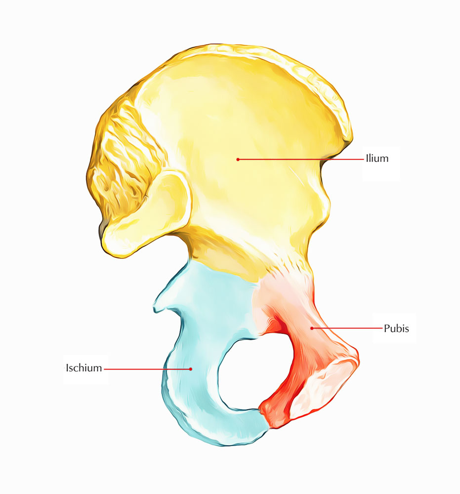
Parts of Hip Bone
Anatomical Position and Side Conclusion
The side of the hip bone is discovered by holding it in such manner that:
- The acetabulum is directed laterally.
- The obturator foramen is located below the acetabulum.
- The flat increased ilium is directed upward, thin pubis is directed anteromedially, and thick ischium is directed posteroinferiorly.
- The pubic tubercle and anterior superior iliac spine be located in the exact same coronal plane.
- The ischial tuberosity takes up the bottom position.
Features and Attachments of Hip Bone
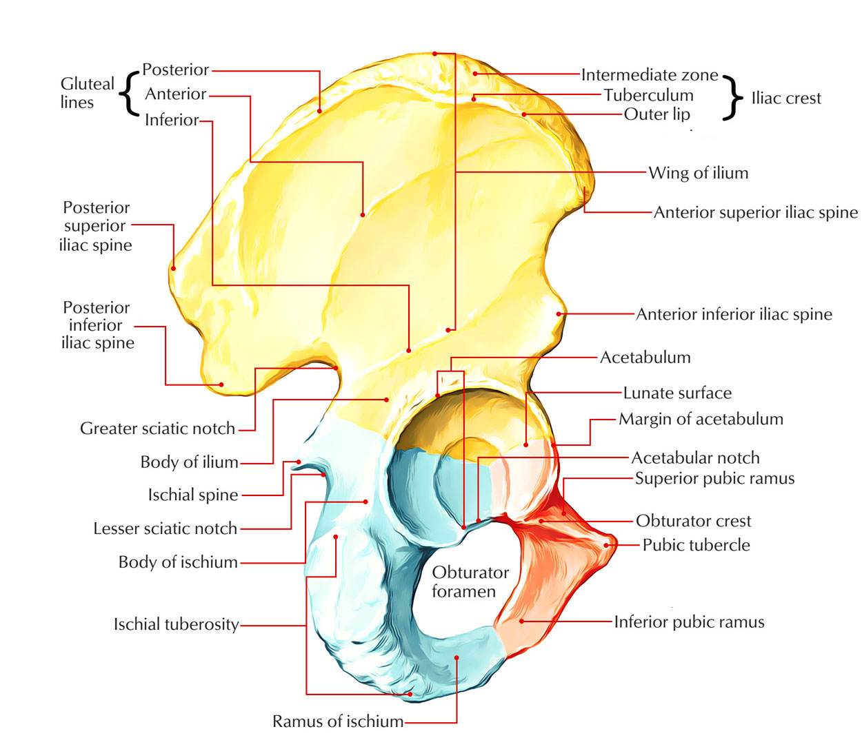
Attachments of Hip Bone
Ilium
It projects upward from the acetabulum as an enlarged fan-shaped plate of bone. It presents these features:
- 2 ends- upper and lower.
- 3 edges- anterior, posterior, and medial.
- 3 surfaces- iliac fossa, gluteal, and sacropelvic.
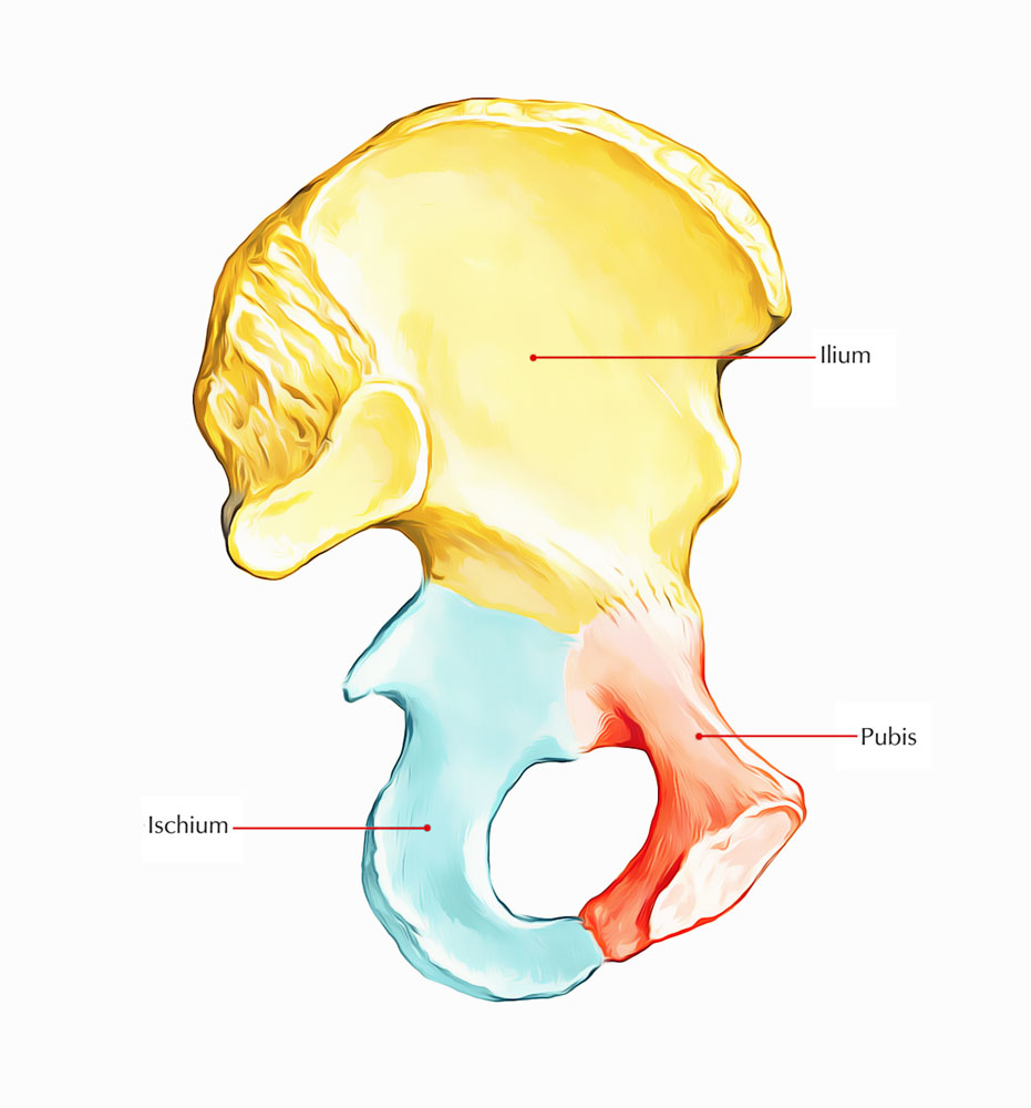
Hip Bone: Ilium
Ends
Upper Ending
1. It’s represented by a curved and thickened border named iliac crest. The maximum point of iliac crest is located at the level of interval between L3 and L4 vertebrae. 2. In the horizontal plane, the iliac crest is concave inward anteriorly and convex inward posteriorly. The anterior and posterior ends of the iliac crest are called anterior superior iliac spine and posterior superior iliac spine, respectively.
- Anterior superior iliac spine gives connection to the lateral end of inguinal ligament above and sartorius below.
- Posterior superior iliac spine gives connection to the sacrotuberous ligament.
3. The iliac crest is split into a bigger ventral section (anterior two-third) and a smaller dorsal section (posterior one-third). Ventral section, presents an outer lip, an intermediate area, and an inner lip.
Outer Lip
It presents tubercle of the iliac crest 5 cm behind the anterior superior iliac spine. It gives connection to these structures:
- Tensor fasciae latae, starts in front of the tubercle of iliac crest.
- External oblique muscle, added in its anterior two-third.
- Latissimus dorsi, comes from just behind itshighest stage.
Intermediate area: It gives origin to the internaloblique muscle along its entire extent.
Inner Lip
1. Its anterior two-third gives connection to transversus abdominis muscle. 2. Its posterior one-third gives connection to quadratus lumborum. Dorsal section is split by a ridge into inner and outer sloping surfaces:
- Outer sloping surface supplies origin to gluteus maximus muscle.
- Inner sloping surface supplies origin to erector spinae muscle.
Lower End
The lower end of ilium creates the upper 2-fifth of acetabulum.
Borders of the Ilium
Anterior Border
- It goes from the anterior superior iliac spine to the acetabulum.
- Its upper half creates a notch and its lower half projects forward as the anterior inferior iliac spine, which gives connection to the straight head of rectus femoris in the upper part and iliofemoral ligament in the lower part.
Posterior Border
- It stretches from the posterior superior iliac spine to the upper end of the posterior border of ischium.
- It presents from above downward, posterior inferior iliac spine, greater sciatic notch, ischial spine, and lesser sciatic notch.
- The lesser and greater sciatic degrees are converted into lesser and greater sciatic foramina by the sacrospinous and sacrotuberous ligaments.
- The dorsal aspect of ischial spine is crossed from lateral to medial side by 3 structures:
- Pudendal nerve
- Internal pudendal vessels
- Nerve to obturator internus
Mnemonic: PIN structures.
Medial Border
- It goes downward, forward, and medially from the iliac crest to the iliopubic eminence
- It divides the iliac fossa from the sacropelvic surface
- Lower one-third of the medial border creates a rounded arcuate line
Surfaces of The Ilium Iliac Fossa
- It’s a shallow concave surface on the inner aspect of ilium, below the iliac crest, and in front of the medial border
- Iliacus muscle originates from its upper two-third
Gluteal Surface
- It’s the outer aspect of the ilium that is convex in front and concave behind.
- It’s split into 4 regions by 3 gluteal lines: posterior, anterior, and inferior. All these lines radiate from the upper margin of the greater sciatic notch. The posterior gluteal line meets the dorsal section of the iliac crest, anterior gluteal line meets the iliac crest near the tubercle of the iliac crest, and inferior gluteal line meets the upper end of anterior inferior iliac spine.
- The gluteal surface gives origin to 4 muscles:
- Gluteus maximus, behind the posterior gluteal line.
- Gluteus medius, between the anterior and the posterior gluteal lines.
- Gluteus medius, between the anterior and the inferior gluteal lines.
- Reflected head of rectus femoris, between the inferior gluteal line and upper margin of acetabulum.
Sacropelvic Surface
It’s situated behind the medial border, on the inner side of ilium. It’s split into 2 parts- sacral part dorsally and pelvic part ventrally. Sacral part of the sacropelvic surface:
- The sacral part presents the tuberosity of the ilium behind and the auricular surface in front.
- The iliac tuberosity is a rough area below the dorsal part of iliac crest and gives connection to 3 ligaments. From behind forwards these are:
- iliolumbar, (b) dorsal sacroiliac, and (c) interosseous sacroiliac.
- The auricular surface is an ear-shaped articular surface and articulates with all the corresponding outermost layer of the sacrum to create a sacroiliac joint.
Pelvic part of the sacropelvic surface:
- It’s smooth and constant with the pelvic outermost layer of the ischium to create the lateral wall of lesser pelvis.
- It presents pre-auricular sulcus just in front of auricular surface, along the lateral border of greater sciatic notch.
- The Majority of this surface gives origin to obturator internus.
Ischium
It’s the thick posteroinferior part of hip bone and is composed of a body and a ramus.

Hip Bone: Ischium
Body
It’s really thick and is located below and posterior to the acetabulum. It presents 2 ends – upper and lower; 3 edges – anterior, posterior, and lateral; and 3 surfaces- femoral, dorsal, and pelvic.
Ends
Upper End
It creates posteroinferior two-fifth of the acetabulum, where it unites with the ilium and pubis.
Lower End
- It creates the ischial tuberosit and produces ramus which creates an acute angle with body.
- Ischial tuberosity is rough and split by a horizontal ridge into an upper quadrilateral area and lower triangular area.
- The upper quadrilateral area is subdivided by an oblique ridge into upper lateral and lower medial parts.
- The lower triangular area is subdivided by a longitudinal ridge into outer and inner parts.
- The ischial tuberosity gives origin to 4 muscles:
- Semimembranosus originates from the lateral part of the upper quadrangular area.
- Semitendinosus together with long head of biceps femoris originates from the medial part of the upper quadrilateral area.
- Adductor magnus (ischial head), originates from the outer part of lower triangular area.
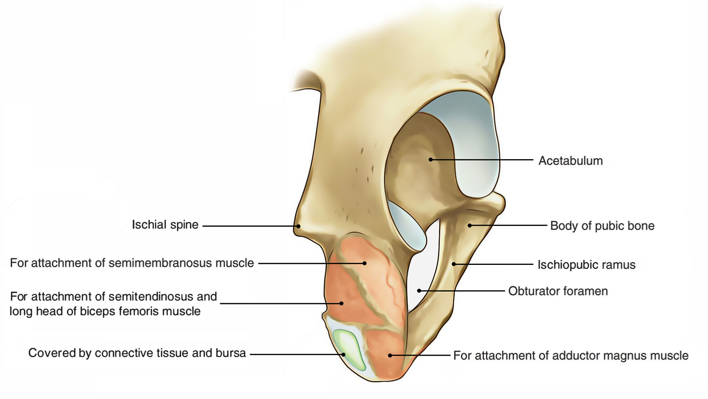
Hip Bone: Ends of Ischium
- The inner part of the lower triangular area is covered by fibrofatty tissue and carries the body weight in sitting position.
- Medial margin of the ischial tuberosity is sharp and gives connection to the lower end of sacrotuberous ligament.
Borders
Anterior Border
It creates the posterior margin of the obturator foramen and provides connection to the obturator membrane.
Posterior Border
- It’s continuous above with the posterior border of ilium and ends below the upper end of ischial tuberosity.
- It presents a triangular projection referred to as ischial spine. The tip of spine gives connection to the sacrospinous ligament, and levator ani and coccygeus muscles originate from its pelvic surface.
- Ischial spine creates the lower boundary of greater sciatic notch and the lower boundary of lesser sciatic notch.
- The gemellus superior and gemellus inferior muscles originate from the upper and lower ends of the lesser sciatic notch, respectively.
Lateral Border
- It creates the lateral margin of the ischial tuberosity.
- Its upper part is notched close to the lower margin of the acetabulum and lodges the tendon of obturator externus.
Surfaces
Femoral Surface
- It’s pointed forwards and laterally, and is situated between the anterior and lateral edges.
- Obturator externus appears from this surface along the obturator foramen.
- Quadratus femoris appears from this surface along the lateral border of the ischial tuberosity.
Dorsal Surface
- It’s continuous above with the gluteal surface of ilium.
- It’s split by a transverse groove into the upper and lower parts.
- Tendon of obturator internus together with 2 gemelli traverses via the groove between the upper and lower parts.
Pelvic Surface
It’s smooth and is located between the anterior and posterior edges. It provides origin to the obturator internus.
Ramus of the Ischium
It appears from the lower part of the body and runs upward, forwards, and medially, and meets with all the inferior ramus pubis to create conjoint ischiopubic ramus.
Conjoint Ischiopubic Ramus
The conjoint ischiopubic ramus presents 2 edges, upper and lower, and 2 surfaces – outer and inner. Upper border creates the lower margin of obturator foramen and gives connection to the obturator membrane. Lower border:
- It creates the lateral boundary of the subpubic angle.
- It’s everted. The eversion is more marked in men than females because of connection of crus of penis.
Outer surface is concave and gives linear connection to 4 muscles. From above downward these are:
- Obturator externus.
- Adductor magnus.
- Adductor brevis.
- Gracilis.
Inner surface is convex and subdivided by the upper and lower oblique bony ridges into 3 regions- upper, middle, and lower.
- Upper ridge gives connection to the superior fascia of urogenital diaphragm.
- Lower ridge gives connection to the perineal membrane (inferior fascia of urogenital diaphragm) and falciform process of sacrotuberous ligament.
- Upper area gives origin to the obturator internus.
- Middle area gives attachments to the sphincter urethrae and deep transversus perinei muscles.
- Lower area gives attachments from before backward to crus of penis, clitoris, ischiocavernosus, and superficial transversus perinei muscles.
Pubis (Pubic Bone/Os Pubis)
It’s the anteroinferior part of the hip bone and is composed of 3 parts: body, superior ramus, and inferior ramus.

Hip Bone: Pubis
Body
It’s quadrilateral in outline and flattened anteroposteriorly. It presents these features: pubic crest, pubic tubercle, and 3 surfaces (anterior, posterior, and medial).
Pubic Crest and Pubic Tubercle
The pubic crest is the thickened upper border of the body of pubis and its lateral end projects forward to create tubercle referred to as pubic tubercle.
Surfaces
Anterior Surface
- It’s pointed downward, forward, and laterally.
- It gives attachments to the subsequent 4 muscles:
- Adductor longus originates by a rounded tendon from the angle between the pubic crest and pubic symphysis.
- Gracilis originates from its lower part and goes farther on the ischiopubic ramus.
- Adductor brevis appears lateral to gracilis and expands farther on the ischiopubic ramus.
Posterior Surface
- It’s smooth and creates the anterior wall of bony pelvis, therefore also referred to as pelvic surface.
- It’s pointed upward and backwards, and is linked to the urinary bladder.
Medial surface (symphyseal surface): It articulates together with the medial surface of the opposite pubic bone to create secondary cartilaginous joint named symphysis pubis.
Superior Ramus of Pubis
- It originates from the superolateral angle of the body of pubis and stretches laterally above the obturator foramen and ends at iliopubic eminence where it unsites with the ilium. It’s triangular in cross section and presents 3 edges- posterior (pectineal line), anterior (obturator crest), and inferior; and 3 surfaces- pectineal, pelvic, and obturator.
Borders
Pectineal Line (Posterior Border)
- It’s also named pecten pubis.
- It stretches laterally as a sharp line from the pubic tubercle to the posterior part of iliopubic eminence where it becomes constant with arcuate line.
- Pectineal line gives connection to a large number of structures:
a) Medial part from before backwards supply attachments to:
- Lacunar ligament
- Represented part of inguinal ligament
- Conjoint tendon
- Fascia transversalis
b) Lateral part from behind forwards gives connection to:
- Pectineal ligament (ligament of Cooper)
- Pectineus muscle
- Pectineus fascia
- Psoas minor (if present)
Obturator Crest (Anterior Border)
It goes from the pubic tubercle to the acetabular margin near the acetabular notch. Inferior border: It creates the upper margin of obturator foramen and gives connection to the obturator membrane making a gap for the obturator canal.
Surfaces
Pectineal Surface
- It’s a triangular area between the obturator crest and the pectineal line.
- It goes from the pubic tubercle to iliopubic eminence.
- Pectineus muscle originates from the upper part of the surface.
Pelvic Surface
It is located between the pectineal line and inferior border of the superior ramus and continues medially with the pelvic outermost layer of the body of pubis. It’s featureless and covered by the peritoneum. The pelvic surface of the superior ramus of pubis is crossed subperitoneally by the obliterated umbilical artery.
Obturator Surface
- It’s situated between the obturator crest and the inferior border.
- Obturator nerve and vessels traverse the obturator groove observed on this particular surface.
Inferior Ramus of Pubis
It goes downward and laterally from the lower part of the body of pubis to join the ramus of ischium and create the conjoint ischiopubic ramus.
Obturator Foramen
- It’s a large gap in the lower part of the hip bone situated between the pubis and ischium.
- It’s oval and bigger in males and triangular and smaller in females.
- Obturator membrane is connected along to its margin with the exception of where it bridges the obturator groove to convert it into obturator canal.
- The obturator canal carries the obturator nerves and vessels.
Acetabulum
It’s a large cup-shaped cavity on the outer outermost layer of the middle constricted part of the hip bone.
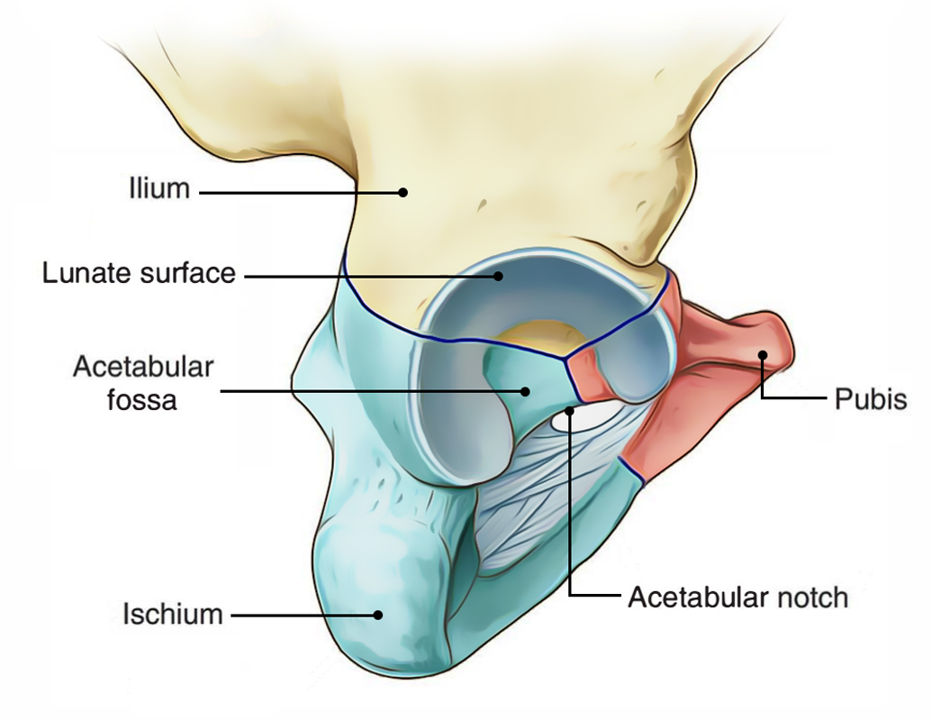
Hip Bone: Acetabulum
It’s pointed laterally and somewhat anteroinferiorly. All the 3 parts of the hip bone lead in its formation as follows:
- Pubis creates its anterior one-fifth.
- Ischium creates its posterior two-fifth.
- Ilium creates its superior two-fifth.
- Its margin is deficient inferiorly to create the acetabular notch, that is bridged by the transverse acetabular ligament, and the notch is converted into acetabular foramen.
- The acetabulum gets the head of femur to create the hip joint.
- The fibrocartilaginous rim, the acetabular labrum, is connected to the margins of the acetabulum.
- The acetabulum presents a horseshoe-shaped articular surface, the lunate surface, in its periphery and non-articular area in its central part, the acetabular fossa. The acetabular fossa is filled up with the pad of fat and the lunate surface is covered by a plate of hyaline cartilage.
Differences Between the Male and Female Hip Bones.
| Features | Female | Male |
|---|---|---|
| Greater sciatic notch | Wider (75°) | Narrower (< 50°) |
| Ischial spine | Not inverted | Inverted |
| Ischiopubic ramus | Thin and not everted | Thick and everted |
| Obturator foramen | Triangular | Oval |
| Acetabular diameter | Less than 5 cm | More than 5 cm |
| Distance between the pubic tubercle and | Less than transverse diameter of | Equal to the transverse diameter of |
| anterior acetabular margin | acetabulum | acetabulum |
| Pre-auricular sulcus | More conspicuous | Less conspicuous |
| Iliac fossa | Shallower | Deeper |
Ossification
The hip bone ossifies in cartilage by 3 primary centers and 8 secondary centers. Ossification of the hip bone
| Centres | Age of appearance | Age of fusion |
|---|---|---|
| Primary centres 1. 1 for ilium 2nd month of IUL 2. 1 for ischium 3rd month of IUL 18th year 3. 1 for pubis 4th month of IUL | ||
| Secondary centres 1. 2 for iliac crest 2. 2 for Y-shaped acetabular cartilage 3. 1 for ischial tuberosity | Puberty | 20-25 years (except acetabular cartilage which ossifies at the age of 17 years) |
Clinical Significance of Hip Bone
There are 2 typical methods of fracturing the hip bone:
- Direct injury to the hip bone, for instance from an automobile crash.
- Forces transferred from the lower limb, for instance a heavy fall on the feet.
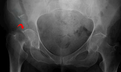
Hip Bone Fracture
Fractures frequently happen at the weaker points of the hip bone. These are the pubic rami, the acetabulum or around the sacroiliac joint. A common problem of hip bone fractures is soft tissue injury. Particularly, the bladder and urethra have high threat of damage.
Test Your Knowledge
Hip Bone

 (55 votes, average: 4.56 out of 5)