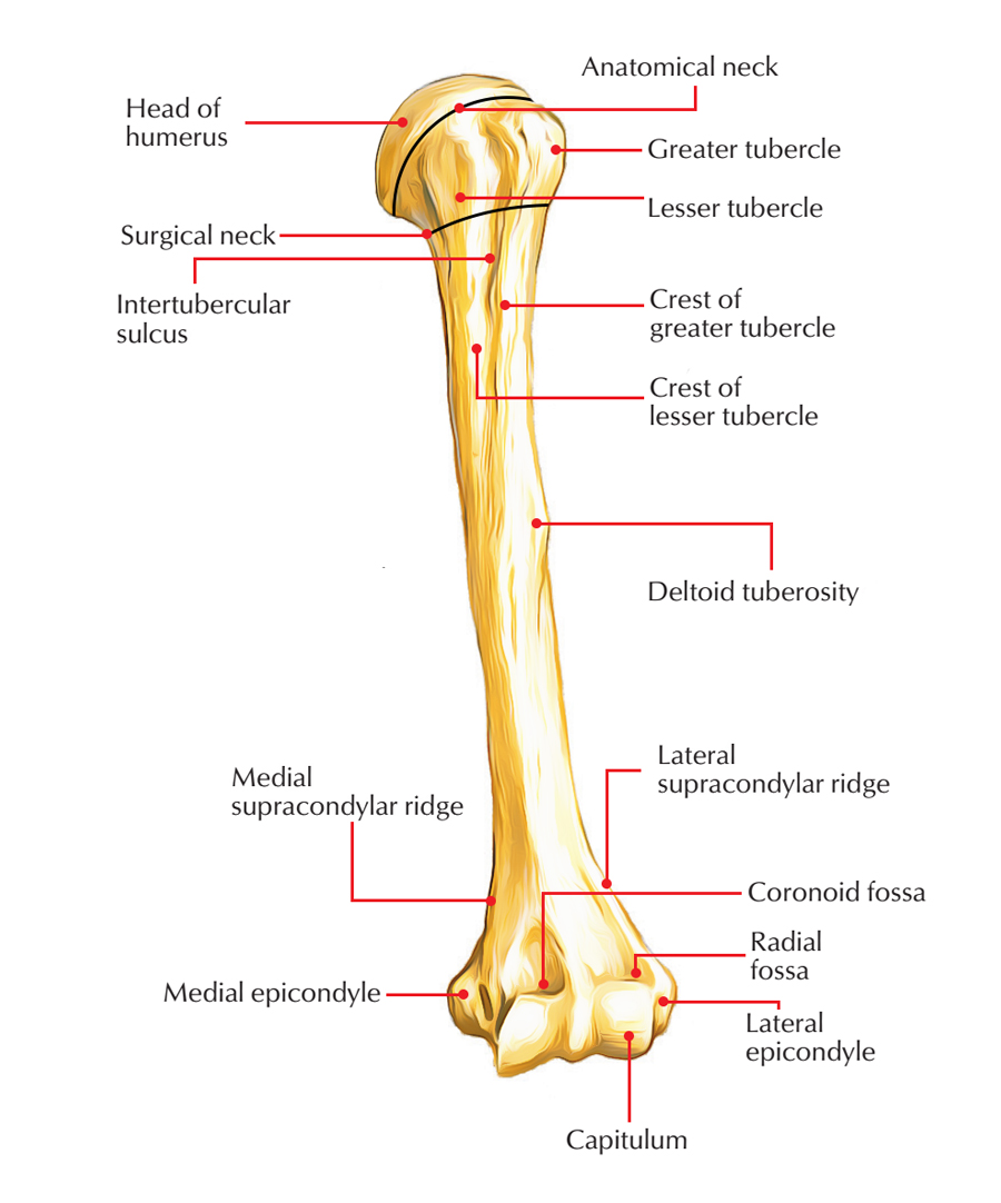
The Humerus is referred to as the bone of the arm and sometimes commonly referred to as the funny bone. It is the longest and also strongest bone of the upper limb. Many muscles which manipulate the arm, at the forearm, at the elbow and the shoulder are anchored to the humerus bone. The region articulates forming part of the shoulder joint. The humerus is the basis to which muscles insert, like the pectoralis major, the deltoid, and others.
Parts of The Humerus Bone
The humerus is a long bone (Based on the types of bones). It is composed of three components: upper end, a lower end, and a shaft.

Humerus Bone
Upper End
The upper end of humerus provides the following 5 features:
- Head
- Neck
- Greater tubercle
- Lesser tubercle
- Intertubercular sulcus
The head is smooth and rounded, and forms less than a fraction of a sphere. It is directed medially backwards and upwards. It articulates along with the glenoid cavity of the scapula to create the glenohumeral (shoulder) joint.
Lower End
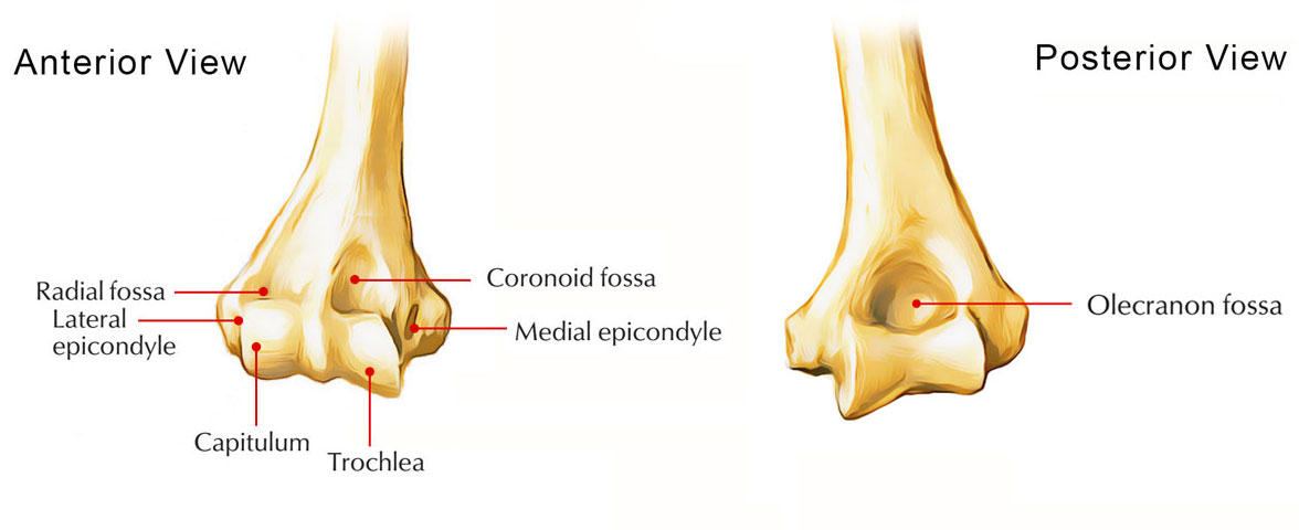
The lower end of the humerus provides the following 7 features:
- Capitulum, a lateral rounded convex projection.
- Trochlea, a medial pulley-shaped structure.
- Radial fossa, a small fossa above the capitulum.
- Coronoid fossa, a small fossa above the trochlea.
- Medial epicondyle, a prominent projection on the medial side.
- Lateral epicondyle, a prominent projection on the lateral side but less than the medial epicondyle.
- Olecranon fossa, a big, deep hollow on the posterior aspect above the trochlea.
Shaft
The shaft is a long part of bone extending between its upper and lower ends. The shaft of the humerus is cylindrical in the upper half but flattened anteroposteriorly in the lower half.
Anatomical Position And Side Determination of The Humerus Bone
The side of the humerus can be identified by holding it vertically in such a way that:
- The rounded head at the upper end faces medially, backwards and upwards.
- The lesser tubercle, greater tubercle, and vertical groove (intertubercular groove) at the upper end faces anteriorly.
- The olecranon fossa on the lower flattened end faces posteriorly.
Features and Attachments of The Humerus Bone
Upper End

Upper End of Humerus
The features of the upper end of the humerus are as follows:
Head
- The head of the humerus is smooth, rounded and makes one-third of a sphere.
- It is encompassed by an articular hyaline cartilage, which in turn is thicker in the core and thinner at the periphery.
Neck
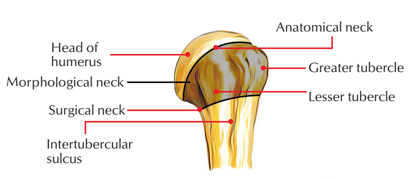
The humerus has three necks:
Anatomical Neck
- The anatomical neck of the humerus is constriction at the margins of the rounded head.
- It provides connection to the capsular ligament of the shoulder joint, apart from – superiorly in which the capsule is deficient, for the passage of tendon of long head of biceps brachii, medially the capsule extends down from the anatomical neck to the shaft for around 1-2 cm
Surgical Neck
- The surgical neck of the humerus is a short constriction in the upper end of the shaft below the greater and lesser tubercles/below the epiphyseal line.
- It is related to the axillary nerve and posterior and anterior circumflex humeral vessels.
- It is one of the most important function of the proximal end of the humerus due to the fact that it is weaker than the more proximal regions of the bone, for this reason, it is one of the sites in which the humerus commonly fractures causing damage of associated nerves and vessels.
Morphological Neck
- The morphological neck of the humerus is the junction between diaphysis and epiphysis.
- It is represented by an epiphyseal line in the adult bone.
- It is a true junction of the head with the shaft.
Greater Tubercle
- The greater tubercle of the humerus is one of the most lateral components of the proximal end of the humerus.
- Its posterosuperior aspect carries three flattened facet-like impressions: upper, middle, and lower, that provide connection to supraspinatus, infraspinatus, and teres minor muscles, respectively.
Lesser tubercle
- The lesser tubercle of the humerus is minor elevation on the front of the upper end of the humerus, just above the surgical neck.
- It supplies connection to subscapularis muscle.
Intertubercular Sulcus/Bicipital Groove
- The Intertubercular Sulcus of the humerus is a vertical groove between lesser and greater tubercles.
- It consists of (a) long head of biceps, wrapped in the synovial sheath and (b) ascending branch of the anterior circumflex humeral artery.
- Three muscles are connected in the region of this groove:
- Pectoralis major on the lateral lip of the groove.
- Teres major on the medial lip of the groove.
- Latissimus dorsi in the floor of the groove.
Mnemonic: Lady between 2 Majors. The ‘L’ of lady stands for latissimus dorsi and ‘2M’ stands for pectoralis major and teres major.
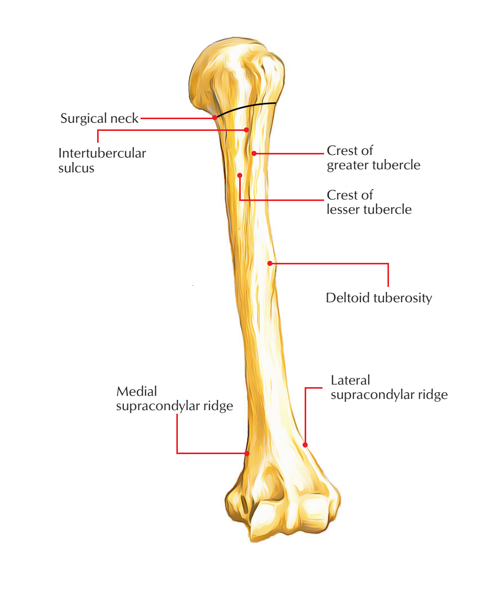
Shaft
The upper part of the humerus is connected with the shaft which is cylindrical. The shaft then is connected to the lower part of the humerus which is triangular in shape when viewed in cross-section. The shaft of the humerus has three borders and three surfaces.

Shaft of Humerus
Borders
Anterior Border
It begins with the lateral lip of the intertubercular sulcus, and extends down to the anterior margin of the deltoid tuberosity and end up being smooth and rounded in the lower half, wherein it ends in the radial fossa.
Medial Border
- It extends from the medial lip of the intertubercular sulcus down to the medial epicondyle. Its lower part is sharp and called medial supracondylar ridge. This ridge offers a connection to the medial intermuscular septum.
- A rough strip on the middle of this particular border offers insertion to the coracobrachialis muscle.
- A narrow area above the medial epicondyle offers origin to the humeral head of the pronator teres.
Lateral Border
- Its upper part is indistinct while its lower part is prominent where it makes up the lateral supracondylar ridge. Above the lateral supracondylar ridge, it is ill-defined nevertheless traceable to the posterior part of the greater tubercle.
- About its middle, this border is crossed by the radial groove from behind.
- The lower component of this border, lateral supracondylar ridge, provides attachment to the lateral intermuscular septum.
Surfaces
Anterolateral Surface
- It is located between the anterior and lateral borders.
- A little above the middle, this surface presents a characteristic V-shaped tuberosity-the deltoid tuberosity which provides insertion to the deltoid muscle.
Anteromedial Surface
- It is located between the anterior and medial borders.
- The upper part of this surface forms the floor of the intertubercular sulcus.
- About its middle and close to the medial border, it presents a nutrient foramen directed downwards.
Posterior Surface
- It is located between the medial and lateral borders.
- In the upper one-third of this surface, generally there is an oblique ridge directed downwards and laterally. This ridge offers an origin to the lateral head of the triceps brachii.
- Below and medial to the ridge, is the radial/spiral groove, which lodges radial nerve and profunda brachii vessels.
- The entire posterior surface below the spiral groove offers an origin to the medial head of the triceps brachii.
Lower End

Lower End of Humerus
The lower end of the humerus is flattened from before backwards and expanded from side to side. It contains the following features:
- The capitulum (rounded convex projection laterally) articulates along with the head of the radius.
- The trochlea (pulley-shaped projection medially) articulates with the trochlear notch of the ulna.
- The ulnar nerve is associated with the posterior surface of the medial epicondyle.
- The anterior surface of the medial epicondyle offers an area for common flexor origin of the superficial flexors of the forearm.
- The anterolateral part of lateral epicondyle provides an area for common extensor origin.
- The posterior surface of lateral epicondyle provides origin to anconeus muscle.
- The coronoid process of ulna fits into coronoid fossa (above the trochlea) during full flexion of the elbow.
- The head of the radius fits into radial fossa (above capitulum) during full flexion of the elbow.
- The olecranon process of ulna fits into olecranon fossa (on posterior aspect above the trochlea) during full extension of the elbow.
- The capsule of the elbow joint is connected above the coronoid and radial fossae anteriorly and above the olecranon fossa posteriorly. In some cases, a bony bar between the coronoid and olecranon fossae is perforated and forms supratrochlear foramen.
- Supratrochlear process: It is a small hook-like bony process, which may sometimes grow from the anteromedial surface, about 5 cm above the lower end of the humerus. A fibrous band named Struthers’ ligament stretches from it to the medial epicondyle. In such cases, the brachial artery along with median nerve deviates from their normal course and pass through the foramen thereby formed.
- The angle of humeral torsion: It is an angle formed by the superimposition of the long axis of the upper and lower articular surfaces of the humerus.
Ossification of The Humerus Bone
The humerus is ossified by the following ossification centres:
- One primary centre for the shaft.
- Three secondary centres for upper end
- Four secondary centres for the lower end.
| Site of look | Time of look | Time of blend | |
|---|---|---|---|
| Shaft | 8th week of IUL | ||
| Upper end | |||
| Head | 1st year | Fuse together at 7th year to form a conjoint upper epiphysis | Accompanies shaft 20th year |
| Greater tubercle | 3rd year | ||
| Lesser tubercle | 5th year | ||
| Lower end | |||
| Capitulum and lateral flange of trochlea | 2nd year | 1 Fuse together at 14th year to form most 1 of the lower epiphysis | Accompanies shaft 16-17th year |
| Medial part of trochlea | 10th year | ||
| Lateral epicondyle | 12th year | ||
| Medial epicondyle | 6th year (form little part of the lower epiphysis) | 18th year | |
Clinical Significance of The Humerus Bone
Nerves directly related to the humerus: The three nerves(axillary, radial, and ulnar) are closely related to the back of humerus as follows:
- Axillary nerve around the surgical neck.
- The radial nerve in the radial/spiral groove.
- Ulnar nerve behind the medial epicondyle.
Therefore, these nerves are often involved in the fracture of the humerus at the above sites:
Common sites of fracture of the humerus: These are (a) surgical neck, (b) shaft, and (c) supracondylar region.
Supracondylar fracture of the humerus: It is triggered by a fall on the outstretched hand and generally takes place in young age. Clinically it provides as the backward displacement of the lower fragment with an unduly prominent elbow, on the other hand, the three bony points of the elbow (viz. medial epicondyle, lateral epicondyle, and olecranon process) form the usual equilateral triangle since the olecranon process always moves with the lower fragment. This fracture may induce injury to median nerve and brachial artery. The injury to the brachial artery may cause Volkmann’s ischemic contracture and myositis ossificans.
Non-union of fracture of the humerus is common, if the fracture occurs at the junction of its upper one-third and middle one-third as a result of poor blood supply.
The median nerve is most commonly associated with the supracondylar fracture of the humerus.
Test Your Knowledge
humerus

 (84 votes, average: 4.51 out of 5)