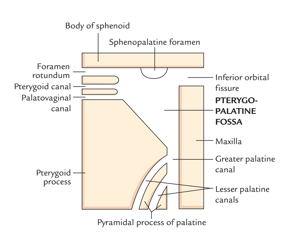
The inferior orbital fissure a.k.a. sphenomaxillary fissure is a longitudinal opening alongside the posterior two-thirds of the bony cavity, located posterolaterally towards the orbital floor dividing the lateral wall of the orbit via its floor.

Inferior Orbital Fissure
Structure
The inferior orbital fissure is thinner centrally compared to the extremities. It is broad in its anterolateral terminal where it is finished by the zygomatic bone.
It conveys:
- Infra-orbital nerve
- Zygomatic nerve
- A branch of the inferior ophthalmic vein and several emissary veins.
- Infra-orbital artery and vein
- Orbital ganglionic branches of the pterygopalatine ganglion.
The fissure is surrounded:
- Posterolaterally – The greater wing of the sphenoid bone.
- Laterally – The zygomatic bone.
- Medially – The sphenoid body and a short section of the palatine bone.
- Anteriorly – The maxilla.
Relations
- Anterior or orbital surface of greater wings of sphenoid bone divides the superior orbital fissure and the inferior orbital fissure.
- Anterior wall of infratemporal fossa and roof of infratemporal fossa is divided by inferior orbital fissure.
- Infratemporal fossa communicates with the orbit via inferior orbital fissure.
- It transmits the:
- Infra-orbital nerve
- Zygomatic nerve
- A division of the inferior ophthalmic vein along with several emissary veins.
- infra-orbital artery and vein
- Orbital ganglionic branches of the pterygopalatine ganglion.
- The fissure is surrounded:
- Posterolaterally – The greater wing of the sphenoid bone.
- Laterally – The zygomatic bone.
- Medially – The sphenoid body and a small section of the palatine bone.
- Anteriorly – The maxilla.
Clinical Significance
Inferior orbital fissure enables connection amongst:
- Posteriorly – Orbit and the pterygopalatine fossa.
- Medially – Orbit and the infratemporal fossa.
- Posterolaterally – Orbit and the temporal fossa.
Periorbital Muscle
Also, the inferior orbital fissure comprises smooth muscle fibers of the periorbital muscle. This muscle belongs to orbital connective tissue system and functions both as an additional muscle:
- For lacrimal function.
- For protruding the eye.
It creates a bridge above the inferior orbital fissure and pterygopalatine fossa, Infratemporal fossa and temporal fossa are separated from the orbit through it. This muscle must not be mixed up with Muller’s muscle which attaches within the tarsal plate of the upper eyelid.

 (53 votes, average: 4.59 out of 5)