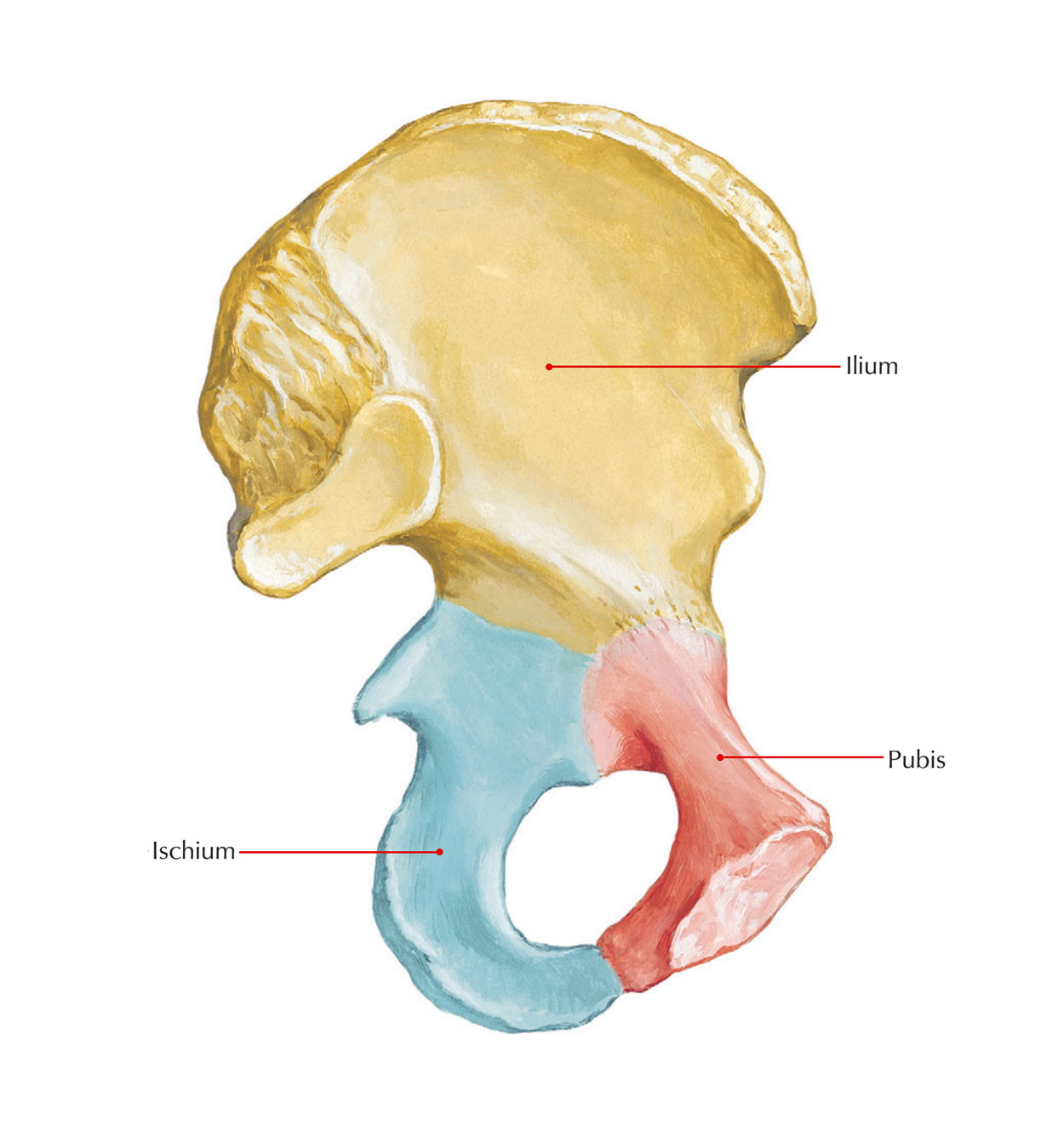
The ischium is located laterally and interiorly to both of the ilium as well as the pubis and creates the posteroinferior part of the os coxa. When an individual is sitting up upright, a person sits on the ischial tuberosities and instead of the legs this bone takes the weight of the body via the sacroiliac joints. It assists significantly in the creation of the acetabulum and its superior border creates the lateral and interior margins of the obturator foramen.
- Below the sacroiliac joint, the posterior border of each ilium indents to create a big greater sciatic notch via which pass blood vessels and nerves.
- The lower portion of the greater sciatic notch is created by ischium, which terminates in a big knob, the ischial tuberosity.
- The hamstrings are a group of big muscles on the backside of the thigh are connected to the ischial tuberosities.

Ischium
Body of the Ischium
This is a thick and massive mass of bone that lies below and behind the acetabulum. It has two ends, upper and lower; three borders, anterior, posterior and lateral; and three surfaces, femoral, dorsal and pelvic.
- The upper end forms the posteroinferior two-fifths of the acetabulum. The ischium, ilium and pubis fuse with each other in the acetabulum.
- The lower end forms the ischial tuberosity. It gives off the ramus of the ischium which forms an acute angle with the body.
- The anterior border forms the posterior margin of the obturator foramen.
- The posterior border is continuous above with the posterior border of the ilium. Below, it ends at the upper end of the ischial tuberosity. It forms part of the lower border of ilium. It also forms part of the lower border of the greater sciatic notch. Below the spine the posterior margin shows a projection called the ischial spine. Below the spine the posterior border shows a concavity called the lesser sciatic notch.
- The lateral border forms the lateral margin of the ischial tuberosity, except at the upper end where it is rounded.
- The femoral surface lies between the anterior and lateral borders. The dorsal surface is continuous above with the gluteal surface of the ilium. From above downwards it presents a convex surface adjoining the acetabulum, a wide shallow groove, and the upper part of the ischial tuberosity.
- The ischial tuberosity is divided by a transverse ridge into an upper and a lower area. The upper area is subdivided by an oblique ridge into a superolateral area and an inferomedial area. The lower area is subdivided by a longitudinal ridge into outer and inner area. The lower area is subdivided by a longitudinal ridge into outer and inner area. The pelvic surface is smooth and forms part of the lateral wall of the true pelvis.
Conjoined Ischiopubic Rami
The inferior ramus of the pubis unites with the ramus of the ischium on the medial side of the obturator foramen. The site of union may be marked by a localized thickening. The conjoined rami have (1) two borders, upper and lower, and (2) two surfaces, outer and inner.
The upper border forms part of the margin of the obturator foramen.
The lower border forms the pubic arch along with the corresponding border of the bone of the opposite side.
The inner surface is convex and smooth. It is divided into three areas, upper, middle and lower, by two ridges.
Attachments and Relations of the Ischium
1. The ischial spine provides (a) attachment to the sacrospinous ligament along its margins and (b) origin for the posterior fibres of the levator ani from its pelvic surface. Its dorsal surface is crossed by the internal pudendal vessels and by the nerve to the obturator intemus.
2. The lesser sciatic notch is occupied by the tendon of the obturator intemus. There is a bursa deep to the tendon. The notch is lined by hyaline cartilage. The upper and lower margins of the notch give origin to the superior and inferior gemelli respectively.
3. The femoral surface of the ischium gives origin to (a) the obturator extemus along the margin of the obturator foramen and (b) the quadratus femoris along the lateral border of the upper part of the ischial tuberosity.
4. The dorsal surface of the ischium has the following relationships. The upper convex area is related to the piriformis, the sciatic nerve, and the nerve to the quadratus femoris. The groove transmits the tendon of the obturator intemus.
5. The attachments on the ischial tuberosity are as follows:
- The superolateral area gives origin to the semimembranosus, the inferomedial area to the semitendinosus and the long head of the biceps femoris, and the outer lower area to the adductor magnus.
- The inner lower area is covered with fibrofatty tissue which supports body weight in the sitting position.
- The sharp medial margin of the tuberosity gives attachment to the sacrotuberous ligament. The lateral border of the ischial tuberosity provides attachment to the ischiofemoral ligament, just below the acetabulum.
6. The greater part of the pelvic surface of the ischium gives origin to the obturator intemus. The lower end of this surface forms part of the lateral wall of the ischiorectal fossa lower end of this surface forms part of the lateral wall of the ischiorectal fossa.
7. The attachments on the conjoined ischiopubic rami are as follows:
- The upper border gives attachment to the obturator membrane.
- The lower border provides attachment to the fascia lata, and to the membranous layer of superficial fascia or Colles’ fascia of the perineum.
- The muscles taking origin from the outer surface are:
(i) the obturator externus, near the obturator margin of both rami,
(ii) the adductor brevis, chiefly from the pubic ramus,
(iii) the gracilis, chiefly from the pubic ramus and
(iv) the adductor magnus, chiefly from the ischial ramus. - The attachments on the inner surface are as follows. The upper ridge gives attachment to the upper layer of the urogenital diaphragm. The perineal membrane is attached to the lower ridge. The upper area gives origin to the obturator internus. The middle area gives origin to the sphincter urethrae and to the deep transverse perinei, and is related to the dorsal nerve of the penis, and to the internal pudendal vessels. The lower area provides attachment to the crus penis, and gives origin to the ischiocavemosus and to the superficial transverse perinei.

 (48 votes, average: 4.75 out of 5)