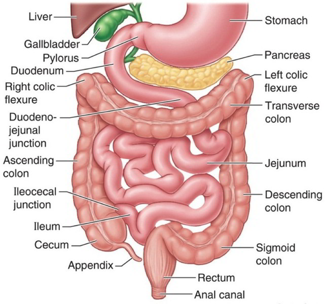
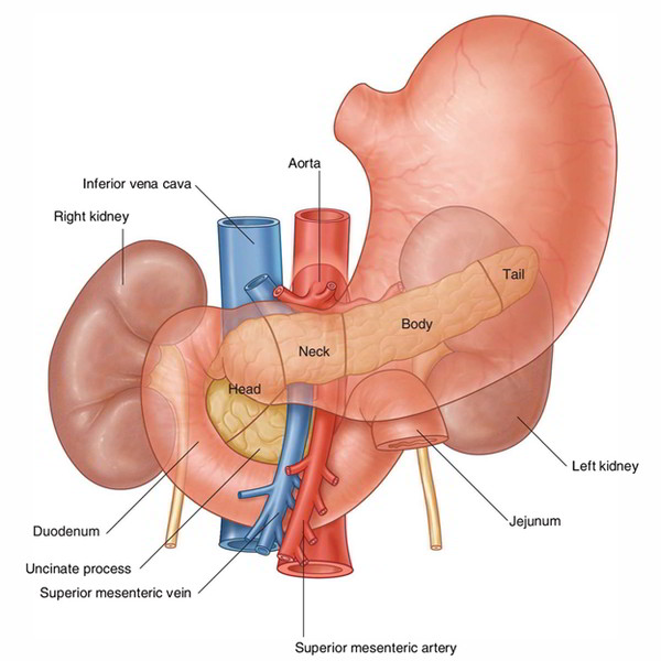
The jejunum is middle part of the small intestine in humans and most higher vertebrates. It lies between duodenum and the ileum. The jejunum arises from the duodenojejunal flexur.
Structure
The wall structure of the small intestine includes four layers. From inside to outside, these are:
- Mucosa
- submucosa
- Muscle layer
- Serosa
Mucosa
The mucosa shows the following three essential functions.
- Larger Surface Area: The surface area of mucous membrane is significantly increased by plicae circulares, villi, and microvilli, for food digestion and absorption of nutrients.
(a) Plicae circulares (or valves of Kerckring): These are rounded folds, which are irreversible and are not eliminated by expansion.
(b) Villi: The surface of circular folds is covered in small (0.5 mm or less in length) finger-like forecasts termed villi.
(c) Microvilli: The surface epithelium enveloping the villi provides microvilli (or striated border).
- The overall surface of mucosa of the small intestine is around 200 sq. meters.
- In every 2 to 4 days the whole epithelial coating of the small intestine is changed.
Intestinal Glands (Crypts of Lieberkuhn)
The epithelium is invaginated in the lamina propria to create intestinal glands, between the bases of villi.
They produce digestive enzymes and mucous.
Lymphatic Follicles
The lamina propria of the mucous membrane consists of two types of lymphatic follicles.
1. Singular lymph follicles: These are dispersed throughout the length of the small intestine and are 1-2 mm in size.
2. Aggregated lymph roots: They create rounded or oval spots called Peyer’s spots. Each Peyer’s spot length differs from 2 to 10 cm and it consists of about 260 singular lymph follicles. Around the antimesenteric border, they exist lengthwise. Peyer’s spots are small, rounded, and fewer in the distal area of the jejunum, and large, oval, and numerous especially in the ileum in its distal part. Peyer’s spots are much larger and most numerous, may extend in the submucosa right after breaching the muscularis mucosa in the distal part of the ileum.
Submucosa
It consists of loose areolar tissue and consists of blood vessels, lymph vessels, and nerve plexus (Meissner’s plexus).
Muscle Layer
Auerbach’s plexus of nerves exists in between these two layers. It is comprised of outer longitudinal and inner rounded layers of smooth muscle.
Serosa
It is created by the visceral peritoneum and is outlined by the simple squamous epithelium.
Mesentery of the Small Intestine
It is a broad fan-shaped fold of the peritoneum, which suspends the small intestine (jejunum and ileum) via the posterior abdominal wall.It has the root (connected margin) as well as free margin (intestinal margin).The root expanding via the duodenojejunal flexure to the ileocaecal junction and is connected to an oblique line throughout the posterior abdominal wall.The duodenojejunal flexure lies to the left side of second lumbar vertebra, whereas ileocaecal junction lies at the right sacroiliac joint.
Arterial Supply
The jejunum and ileum are supplied by the jejunal and ileal branches (12 – 15 in number) of the superior mesenteric artery. They enter the mesentery to reach the intestine and emerge via the left side of the superior mesenteric artery.
Venous Drainage
The veins represent the branches of superior mesenteric artery and drain right into the portal vein, which brings the items of protein and carbohydrates towards the liver.
Lymphatic Drainage
The lymphatics of the small intestine have a rounded route in its wall. The lymph vessels via the small intestine travel through a great deal of mesenteric nodes (lymph nodes found in the mesentery) and finally drain into superior mesenteric nodes present around the origin of superior mesenteric artery.
Nerve Supply
The sympathetic supply is originated from T10-T11 spinal sections via splanchnic nerves and superior mesenteric plexus. The small intestine is innervated by both sympathetic and parasympathetic nerve fibers.


 (47 votes, average: 4.76 out of 5)