A condyle is a round protrusion which is located at the terminals of a bone. It is a portion of a joint and most commonly is an articulation with another bone. It is one of the anatomical markings or features of bones.
Elbow joint
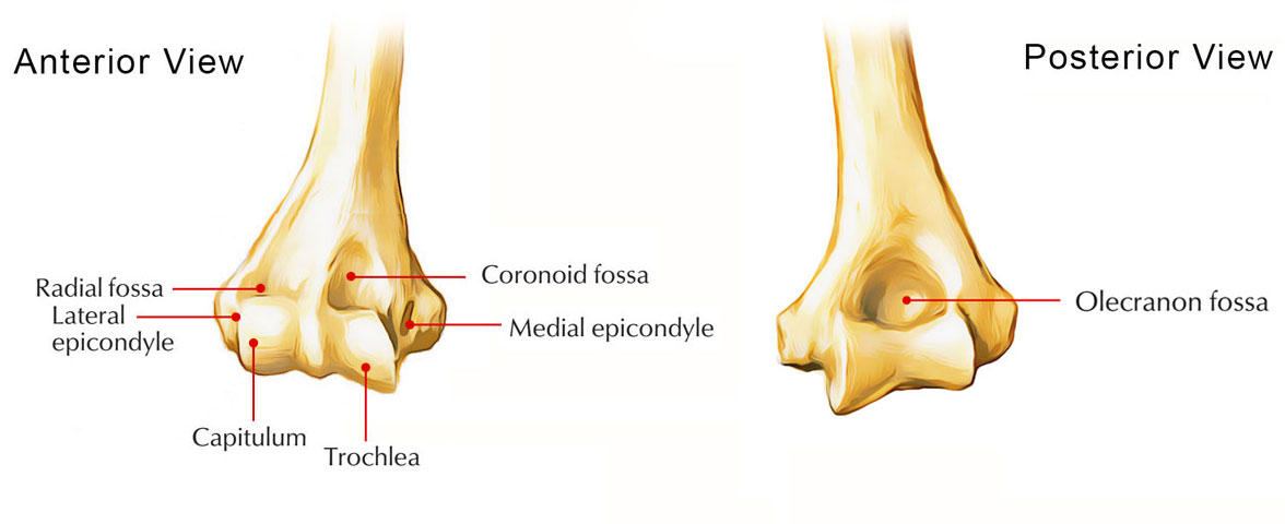
The two articular parts of the condyle connect with the two bones of the forearm,the capitulum and the trochlea.
Capitulum of the Humerus
The capitulum of the humerus acts as a lateral condyle. The capitulum and the radius of the forearm articulate with each other. It is lateral in position and hemispherical in shape, and is not visible when the humerus is seen from the posterior side;it projects anteriorly as well as inferiorly to a certain extent.

Capitulum of the Humerus
Knee Joint
The knee joint contains two separate condyloid joints:
- The medial condyle of the femur connects together with the medial condyle of the tibia
- The lateral condyle of the femur connects together with the lateral condyle of the tibia
The femoral condyles have rounded articular surfaces, while the tibia condyles have flatter articular surfaces, creating small surface areas of interaction among the two bones.
Tibia
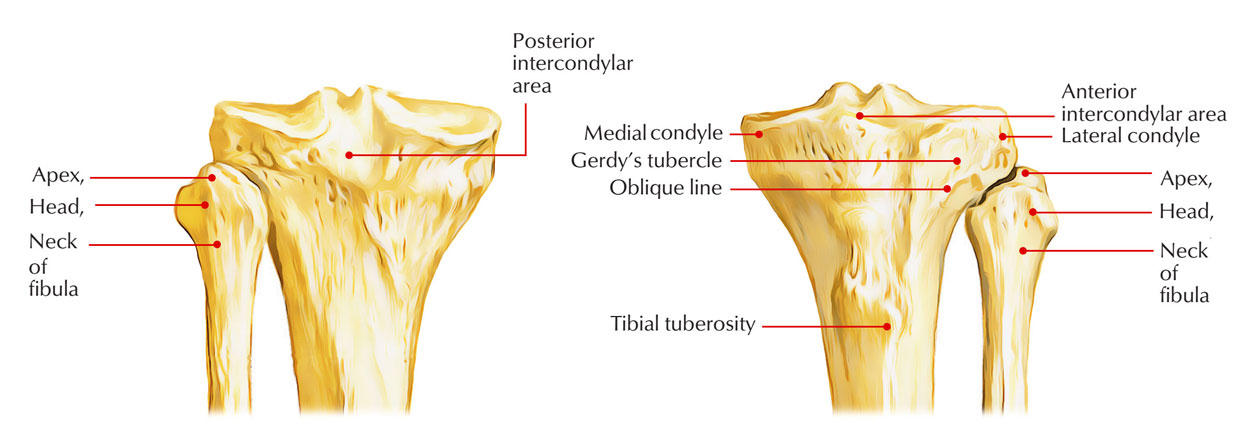
The tibia and the fibula articulate at the two tibiofibular joints. The proximal tibiofibular joint is a plane joint that enables a small amount of gliding, amongst the head of the fibula as well as the posterior portion of the outer side of the lateral condyle of the tibia. The distal tibiofibular joint which pivots slightly is a fibrous joint.
The lateral surface of the distal extension of the tibia is irregular in order to hold the lateral malleolus of the fibula at the distal joint. For proper ankle joint function the slight movement in both tibiofibular joints is necessary however these actions are not visible on the surface.

Lateral Condyle of Tibia
Femur
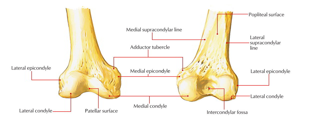
Lateral Condyle of femur is located in lower end of femur and is a major attachment and articulation site. Compared to the medial condyle, the lateral condyle is stouter and stronger,however it is less protuberant.
A protrusion called lateral epicondyle is presented by its lateral surface, which givesconnection to the fibular collateral ligament of the knee joint.

Lateral Condyle of Femur
Origin to the lateral head of gastrocnemius is given by smooth impression above as well as behind the lateral epicondyle. Attachment to popliteus in its anterior part is given by an indentation below and behind the lateral epicondyle.
During full flexion at the knee joint the tendon of popliteus occupies the posterior part of this indentation. The lateral boundary of intercondylar fossa is created by its medial surface.

 (60 votes, average: 4.67 out of 5)