The Medial Compartment of the Thigh is one of the fascial compartments of the thigh and the adductor compartment of the thigh is well developed. Its counterpart in the arm has gotten degeneration during the course of development and is represented only by a feeble coracobrachialis muscle of the flexor compartment of the arm.
Borders
The medial compartment of the thigh is bounded:
- Anteriorly by the anterior intermuscular septum which divides it from the anterior (extensor) compartment of the thigh.
- Posteriorly by an ill defined posterior intermuscular septum which divides it from posterior (flexor) compartment of the thigh. The posterior intermuscular septum is ill defined and incomplete as a result of presence of a composite muscle, the adductor magnus, comprising 2 parts- adductor and flexor (hamstring) belonging to adductor and flexor compartments of the thigh, respectively.
Contents
- Muscles: Adductor longus, adductor brevis, adductor magnus, gracilis, pectineus, and obturator externus.
- Nerve: Obturator nerve.
- Arteries: Profunda femoris artery and obturator artery.
The obturator externus is located deep in this region and is functionally associated with the gluteal region. It rotates the thigh laterally; the main function of others is adduction of the thigh.
Muscles
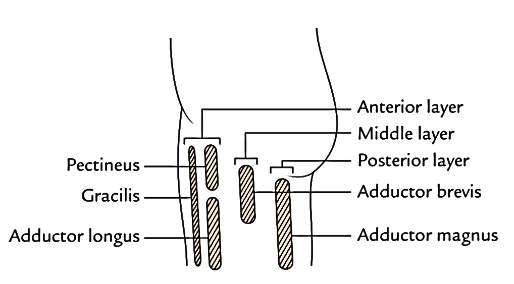
The muscles of the medial compartment of the thigh are ordered into 3 layers. 
- Anterior (first) layer is composed of pectineus, adductor longus, and gracilis.
- Middle (second) layer contains adductor brevis.
- Posterior (third) layer is composed of adductor magnus.
Points to be noticed
- All the muscles of the adductor compartment of the thigh are supplied by the obturator nerve.
- Two muscles of the adductor compartment of thigh, viz., pectineus and adductor magnus, are complex muscles, for this reason they’ve double innervations. The pectineus is supplied by the femoral and obturator nerves while the adductor magnus is supplied by the obturator nerve (posterior section) and tibial part of sciatic nerve.
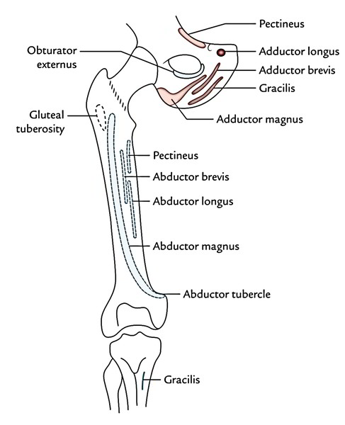
Origin, Insertion, Nerve Supply, and Actions of Muscles
| Muscle | Origin | Insertion | Nerve supply | Actions |
|---|---|---|---|---|
| Pectineus (flat, quadrilateral muscle, composite muscle) | 1. Pecten pubis 2. Upper half of the pectineal surface of the superior ramus of the pubis 3. Fascia covering the pectineus | Line extending from the lesser trochanter to the linea aspera | Femoral nerve and anterior division of the obturator nerve | Adduction of the thigh |
| Adductor longus (triangular muscle, forming the medial part of the floor of the femoral triangle) | Body of the pubis in the angle between the pubic crest and the pubic symphysis | Middle 1/3rd of linea aspera | Obturator nerve (anterior division) | Adduction and medial rotation of thigh |
| Adductor brevis (triangular muscle lying behind the pectineus and adductor longus) | 1. Anterior surface of the body of pubis 2. Outer surface of the inferior ramus of the pubis between the gracilis and the obturator externus | Line extending from the lesser trochanter to the linea aspera, and upper part of the linea aspera itself just lateral to pectineus | Obturator nerve (anterior and posterior divisions) | Adduction of thigh |
| Adductor magnus (large composite) | (a) Hamstring part: inferolateral part of the ischial tuberosity (b) Adductor part: outer part of the ischiopubic ramus | 1. Medial margin of gluteal tuberosity 2. Linea aspera 3. Medial supracondylar line 4. Adductor tubercle | 1.Adductor part by the obturator nerve (posterior division) 2. Hamstring part by the tibial part of sciatic nerve | 1. Adduction and medial rotation of thigh 2. Weak extension of hip joint |
| Gracilis | Medial margin of the lower half of the body of the pubis: adjoining anterior part of inferior ramus of the pubis | Upper part of the medial surface of tibia between the insertions of sartorius (in front) and the semitendinosus (behind) | Obturator nerve (anterior division) | 1. Adduction of thigh 2. Flexion and medial rotation of leg |
x


 (48 votes, average: 4.52 out of 5)