Located at the terminals of a bone, a condyle is a round protrusion, which most commonly is an articulation with another bone and is a portion of a joint. It is among the anatomical or indicating markings or features of bones.
Elbow Joint
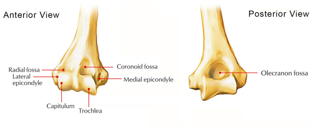
The two articular parts of the condyle connect with the two bones of the forearm, the capitulum as well as the trochlea.
Trochlea of the Humerus
The trochlea and the ulna of the forearm articulate together with each other. It is located medial to the capitulum and is pulley shaped. Compared to the capitulum, it extends over the posterior surface of the bone and its medial edge is more distinct compared its lateral edge.

Medial Condyle of Humerus
Knee Joint
The knee joint comprises of two separate condyloid joints:
- The medial condyle of the femur connects together with the medial condyle of the tibia
- The lateral condyle of the femur connects together with the lateral condyle of the tibia
The femoral condyles have rounded articular surfaces, while the tibia condyles have flatter articular surfaces, creating small surface areas of interaction among the two bones.
Tibia
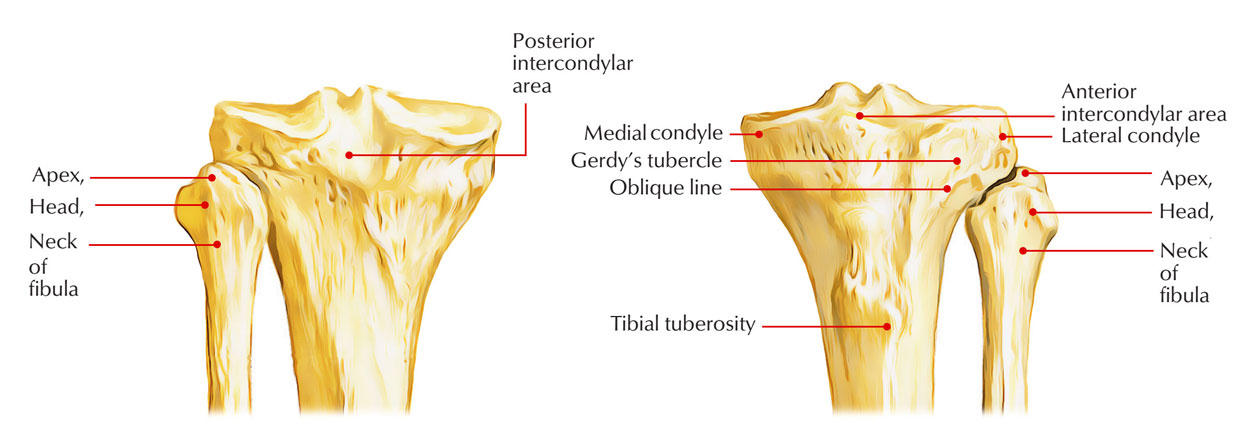
The tibial condyles are thick horizontal discs of bone attached to the top of the tibial shaft. The medial condyle is better reinforced over the shaft of the tibia and it is larger compared to the lateral condyle.
For articulation with the medial condyle of the femur, it has oval superior surface. On the side of the elevated medial intercondylar tubercle, the articular surface spreads laterally.

Medial Condyle of Tibia
Femur
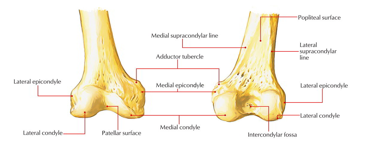
Medial Condyle is located in lower end of femur and is a major attachment and articulation site. Attachment to the upper end of medial collateral ligament is given by its most protuberant point called medial epicondyle.
Insertion to the ischial head of adductor magnus is given by a projection called adductor tubercle that is posterosuperior towards the medial epicondyle. Medial boundary of intercondylar fossa is created by its lateral surface.

Medial Condyle of Femur

 (50 votes, average: 4.72 out of 5)