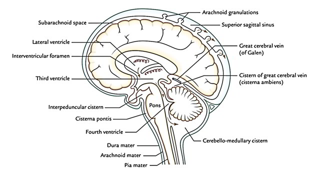
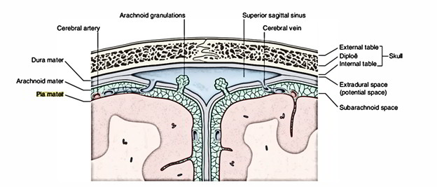
The brain and spinal cord are enclosed inside 3 protective membranes referred to as meninges. From without inward these are as follows: Dura mater, Arachnoid mater and Pia mater.
Originally, the dura mater is mesodermal, on the other hand the arachnoid mater and pia mater are ectodermal in origin (originated from neural crests).
Dura Mater
The cerebral dura mater (also named cranial dura) is a powerful fibrous membrane being composed of 2 layers: outer bargain and inner meningeal layers. With the exception of where they enclose venous sinuses, the 2 layers are fused with every other.
Arachnoid Mater
- The arachnoid mater is an extremely thin, a transparent membrane, which invests the brain loosely and continues as the spinal arachnoid at the foramen magnum, which finishes at the level of second sacral vertebra. It’s closely associated with the internal surface of the dura mater and has precisely the same shape as the dural sac with the exception of where its arachnoid granulations pierce the dura mater.
- The arachnoid mater is split from the dura mater by a capillary space known as the subdural space consisting of a film of fluid. This creates a sliding plane where movement is possible between the dura mater and brain enclosed in the arachnoid and pia mater.
- The subdural space is traversed by cerebral veins on their route to the dural venous sinuses.
Processes of Arachnoid
Arachnoid Villi
All these are great fingerlike processes which originate from the surface of arachnoid. They shove the dura before them and eventually perforate it to project into the dural venous sinuses. They’re covered by specialized mesothelial cells which express the CSF to bloodstream, thereby resulting in the absorption of CSF.
Arachnoid Granulations (Pacchionian Bodies)
Withadvancing age, the arachnoid villi enlarge in size and create pedunculated tufts referred to as arachnoid granulations. Some consider that these are aggregations of arachnoid villi, clumped together, i.e., arachnoid granulations are the large clusters of arachnoid villi. Arachnoid granulations like arachnoid villi are concerned with the absorption of CSF. They project into the venous lacunae of the superior sagittal sinus.
Pia Mater
- Pia mater is a thin diaphanous vascular membrane which closely invests the top layer of the brain. It is adherent to the face of the brain and follows closely the irregularities of its surface.
- All the blood vessels to the brain run in the subarachnoid space on the surface of the pia mater before going into the brain.
- In some specific scenarios, the walls of the cavities of the brain (ventricles) are really thin and created from only an individual layer of its lining epithelium, the ependyma. In such regions, the pia mater being located on its external surface invaginates into ventricular cavities as a series of vascular tufts of capillaries which carry the ependyma before them, thereby creating the choroid plexuses of the brain. The pial component of choroid plexus is referred to as tela choroidea.


 (52 votes, average: 4.81 out of 5)