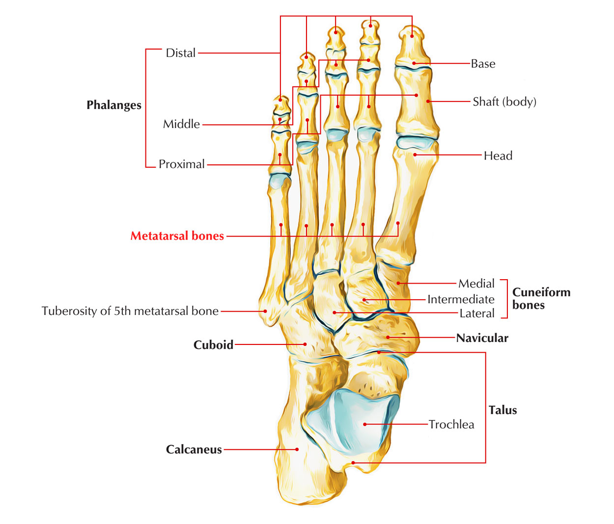
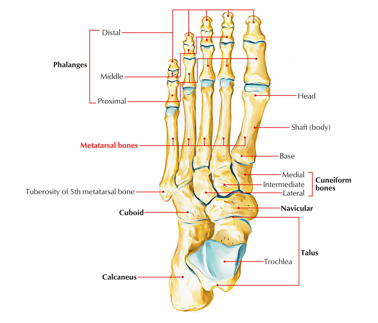
The Metatarsal Bones or Metatarsus are a group of five long bones in the foot. All these are 5 tiny long bones. The 5 metatarsal bones collectively make up the metatarsus.
They can be numbered from medial to lateral sides as first, second, third, fourth, and fifth.
Side Determination
Determination of the side of a metatarsal bone is as follows:
- The bases of the metatarsals are set obliquely so that the deeper edge is directed backwards and laterally.
- The side of the first metatarsal bone can be found out by examining the kidney-shaped facet on the base, the hilum of which is directed laterally.

Metatarsal Bones
Identification
First metatarsal
- It’s the shortest, thickest, and most powerful, and is accommodated for weight transmission.
- Proximal surface of its base presents a kidney shaped articular surface.
Second metatarsal
- It’s the longest metatarsal bone.
- Proximal surface of its base has a triangular concave articular surface.
- The lateral side of the base has two articular facets, a larger dorsal, and a smaller plantar each of which is subdivided into a proximal part for the lateral cuneiform bone and a distal part for the third metatarsal.
- The medial side of the base bears one facet, placed dorsally, for the medial cuneiform.
Third metatarsal
- Proximal surface of its base has a flat triangular articular facet.
- The lateral side of its base has 2 facets while the medial side has 1 facet.
Fourth metatarsal
- The proximal surface of its base has a quadrilateral facet, which articulates with the cuboid.
- The lateral side of base has 1 facet while its medial side has 1 facet split into 2 parts- proximal and distal.
- The medial side of the base has one facet placed dorsally, which is subdivided into a proximal part for the lateral cuneiform and a distal part, for third metatarsal bone.
Fifth metatarsal
- The lateral side of the base has a large tuberosity or styloid process projecting backwards and laterally.
- The medial side of the base has one facet for the fourth metatarsal bone.
- The plantar surface of the base is grooved by the tendon of the abductor digiti minimi.
Features and Attachments
Every metatarsal contains 3 parts: distal end (head), shaft (body), and proximal end (base).

Metatarsal Bones
Head
- It articulates with the base of corresponding proximal phalanx to create the metatarso-phalangeal (MP) joint.
- Every side of the head presents a tubercle dorsally to supply connection to the collateral ligament of MP joint.
Shaft
- Its plantar aspect is concave from before backwards.
- Sides of the shaft supply attachments to the interossei muscles.
- Plantar aspect of the fifth metatarsal supplies origin to the flexor digiti minimi.
Base
Contours of the proximal surfaces of the foundations of the metatarsals and their joints
| Metatarsal | Shape of the articular surface | Articulation |
|---|---|---|
| 1st | Kidney-shaped | Medial cuneiform |
| 2nd | Triangular | Intermediate cuneiform |
| 3rd | Triangular | Lateral cuneiform |
| 4th | Quadrangular | Cuboid |
| 5th | Triangular | Cuboid |
Base of the very first metatarsal gives insertion to:
- Tendon of tibialis anterior inferomedially.
- Tendon of peroneus longus inferolaterally.
Plantar aspects of foundations of middle 3 (second to fourth) metatarsals supply insertion to slides of tibialis posterior and origin to the oblique head of adductor hallucis.
Base of the fifth metatarsal gives these attachments:
- Peroneus brevis is added on the styloid process of base.
- Peroneus tertius is added on the dorsal aspect of the base.
- Flexor digiti minimi brevis comes from the strategytar aspect of the base.
Important Attachments to Metatarsal Bones
- A part of the tibialis anterior is inserted on the medial side of the base of the first metatarsal bone .
- The greater part of the peroneus longus is inserted on a large impression at the inferior angle of the lateral surface of the base of the first metatarsal
- The peroneus brevis is inserted on the dorsal surface of the tuberosity of the fifth metatarsal bone.
- The peroneus tertius is inserted on the medial part of the dorsal surface of the base and the medial border of the shaft of the fifth metatarsal bone.
- The flexor digiti minimi brevis arises from the plantar surface of the base of the fifth metatarsal bone.
- The shaft of metatarsal bones gives origin to interossei.
Ossification
- Every metatarsal ossifies from 2 centers- 1 primary and 1 secondary.
- Primary center for shafts of all the metatarsals appears during 9th week of IUL with the exception of the shaft of first metatarsal which appears during 10th week of IUL.
- Secondary center appears during 3rd to 4th year in the heads of all the metatarsals with the exception of in the very first metatarsal where it appears in the base. All of them join together with the shaft during 17-20 years.
Differences Between Metatarsals And Metacarpals
| Metatarsals | Metacarpals |
|---|---|
| More massive and more strong | Less massive and less strong |
| Head is smaller than base (except first) | Head is larger or equal to the base (except first) |
| Dorsal surface of the shaft presents no flattening | Dorsal surface presents triangular flattening |
| Shaft narrows from the base to the head | Shaft thickens from the base to the head |
| Concave surface of the shaft faces downward | Concave surface of the shaft faces forward |
| Numbered (1 to 5) from medial to lateral side | Numbered (1 to 5) from lateral to medial side |
Clinical Significance
Fracture of the Tuberosity (Styloid Process) of The Fifth Metatarsal
It generally takes place because of forced inversion of the forefoot, when the tendon of peroneus brevis pulls off the styloid process leading to its avulsion. This fracture is also referred to as Jones’ fracture following the name of an orthopaedic surgeon Sir Robert Jones, who himself endured this injury.
March Fracture
It usually appears in the shaft or neck of second and third metatarsals because of competitive protracted march past by the soldiers.

 (50 votes, average: 4.74 out of 5)