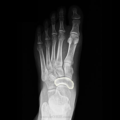
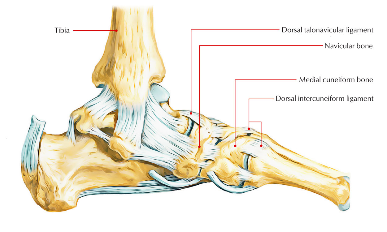
The intermediate tarsal bone on the medial aspect of the foot is the navicular (boat formed). This bone articulates behind with the talus and links in front as well as on the lateral aspect with the distal set of tarsal bones.
One distinguishing characteristic of the navicular is a noticeable curved bulge for the connection of the tibialis posterior tendon that predicts inferiorly on the medial aspect of the plantar surface of the bone.
Surfaces
It is positioned in between the talus posteriorly along with the three cuneiform bones anteriorly. It is formed like a boat and also has 6 surfaces.
Anterior Surface
This surface is entirely artiular and also has three articular aspects, every single triangular fit. The medialmost is the biggest; it’s pinnacle points medially and also links with the medial cuneiform bone. Those for the other two cuneiforms arc also triangular however their peaks point inferiorly.
Posterior Surface
It has a scooped aspect for unification with the head of the talus.
Dorsal Surface
It is completely ligamentous and provides connections to the
• Dorsal talonavicular ligament posteriorly
• Cuncionavicular ligaments anteriorly
• Cubonavicular ligament laterally
Plantar Surface
It is rough and significant by a groove simply lateral to the bulge of the medial surface.
• Groove: Lodges the tendon of the part of the tibialis posterior that continues forwards in the groove and is placed in other tarsal and sonic of the metatarsal bones.
• Surface lateral to the groove: Offers connection to plantar calcaneonav icular (spring) ligament.
Medial Surface
It provides the bulge of the navicular bone in its lower part that gets the insertion of the main part of the tibialis posterior tendon.
Lateral Surface
It is normally non-articular however might in some cases have an aspect for unification with the cuboid bone. The remainder of the surface offers connection to the medial limb (calcaneonavicular part) of the bifurcated ligament.
Unifications
The navicular bone links proximally with the head of the talus bone, laterally with the cuboid bone as well as distally to the lateral, intermediate and medial cuneiform bones.
Development
• Problems of the ossification of the navicular can be common. The navicular is the last bone in the foot to ossify. In ladies, the navicular anlage ossifies in between 18-24 months and in young boys 30-36 months
• The navicular is a boat-shaped bone situated in the leading inner aspect of the foot, simply above the transverse. It assists link the talus, or anklebone, to the cuneiform bones of the foot.
• The dorsalis pedis artery supplies blood to the navicular’s dorsal (upper) aspect, meantime the medial plantar artery provides its plantar (closer to the sole of the foot) area. It is stimulated by the medial plantar and deep fibular nerves.
Clinical importance
Navicular Injuries
• Injury to the navicular, that interrupts either its position or capability to slide over the talus, can significantly hinder foot function.
• Both direct and indirect forces have a function in disturbance of the regular navicular architecture. A direct impact, while uncommon, can trigger avulsions to the circumference or break injury in the dorsal-plantar plane.
• Frequently, indirect forces of central loading possibly straight around the long axis of the foot or obliquely can trigger navicular injury.
• Injury might be sports associated, secondary to a fall through a height, or to the outcome of an automobile collision.
• Because of the cross interaction of the surrounding bony as well as soft tissue constructs, injury to nearby constructs is most likely and should be thoroughly dismissed where a navicular injury is discovered.
• Considerable injury to the cuboid or intcrcunciform constructs can accompany reasonably low-level oblique loads.
Navicular Tension Fracture

Navicular Tension Fracture
Stiff midfoot metatarsus adductus, decreased dorsiflexion, and/or reduced subtalar motion are danger aspects. The condition takes place after recurring longitudinal tension to the bone as it gels compressed in between the talus and cuneiforms. Fracture takes place generally in the center third, reasonably avascular area. This is an unusual fracture and is frequently neglected.
Symptoms and signs
Perilous start of unclear pain at dorsomedial midfoot or medial arch; pain on palpation of navicular bone throughout the dorsal as well as sole; history of weight-bearing pain in a runner or jumper; reduced dorsiflexion due to pain.


 (46 votes, average: 4.79 out of 5)