Neck is a cylindrical part that joins the head to the trunk (Thorax). It mechanically, physically and functionally supports head. The weight, connections and movements of the head are supported by the neck.
Topographical Organization of the Neck
Neck possesses flexiblity and is a passage point of structures like spinal cord, trachea, esophagus, blood and lymphatic vessels, cranial nerves vertebral column etc. all of these structures are vital for life and survival. Like a collar, the investing layer of deep cervical fascia covers the neck. In its course around the neck, it divides to enclose sternocleidomastoid and trapezius muscles. The 2 fascial layers (referred to as pretracheal and prevertebral fasciae) split the neck into anterior and posterior compartments, stretching from the investing layer of deep fascia across the structures inside the neck. 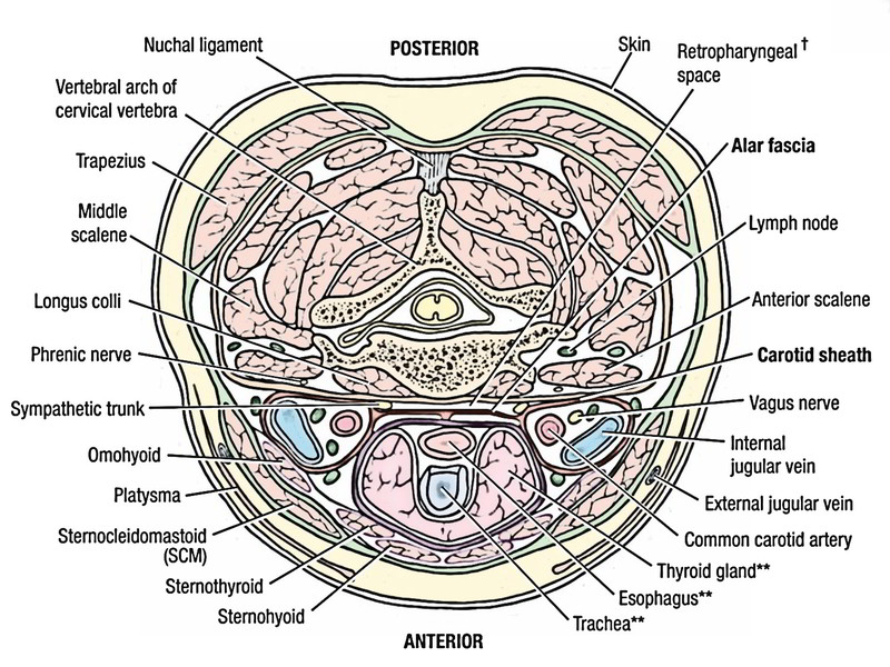
Anterior Compartment
The basic topography of the anterior compartment is simple. In the midline there are 2 tubes: the respiratory tract (larynx and trachea) in front and digestive tract (pharynx and esophagus) behind. The thyroid gland clasps the front and sides of the larynx and trachea and overlaps the carotid tree on each side. These structures are bounded anteriorly by pretracheal fascia, which stretches on either side to unite together with the investing layer of deep cervical fascia deep to sternocleidomastoid. 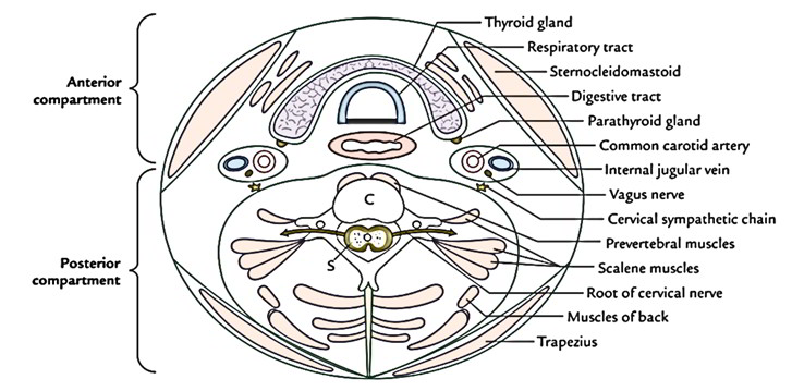
Posterior Compartment
The posterior compartment of neck is composed of cervical part of vertebral column and its surrounding musculature. This musculoskeletal block is bounded by prevertebral fascia, which unifies behind on either side together with the deep fascia enclosing the trapezius muscle. The musculature contains: (a) prevertebral muscles found in front of the cervical column, (b) scalene muscles going between the neck and upper 2 ribs and (c) muscles of the back of the neck. The vertebral canal inside the cervical vertebral column gives passage to the spinal cord. The roots of cervical spinal nerves come out via intervertebral foramina in this region. The ventral rami of the very first 4 cervical nerves create the cervical plexus and ventral rami of the lower 4 cervical nerves together with ventral ramus of T1 create the brachial plexus. The neck, for that reason, is a complex region of the body. The spinal cord, digestive and respiratory tracts and major blood vessels traverse this exceptionally flexible area. The neural structures within the region contain: last 4 cranial nerves and cervical and brachial plexuses. Several organs are also found here. The musculature of neck creates a range of movements in this area. 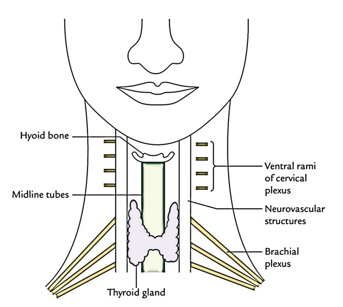
Regions of the Neck
The neck is split into the 4 regions:
- Anterior region.
- Right lateral region.
- Left lateral region.
- Posterior region (nucha).
Anterior Region (Cervix)
The anterior region of the neck includes strap muscles, digestive (pharynx and esophagus) and respiratory (larynx and trachea) tracts, vessels to and from the head, last 4 cranial nerves and thyroid and parathyroid glands. These structures can be easily felt in the anterior region of the neck. In the midline:
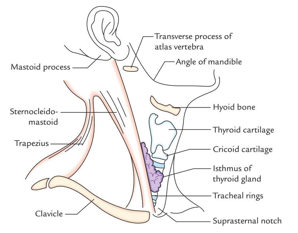
- Hyoid bone: It’s situated in a depression behind and somewhat below the chin and can be easily felt if the neck is somewhat extended. The hyoid bone can be grasped between the thumb and index finger and went from side to side.
- Thyroid cartilage: It’s the most notable characteristic in the anterior region of the neck, especially the anterior angle created by the fusion of its 2 laminae which create the laryngeal prominence. It’s noticeable in men and named Adams apple on the other hand in females it’s not generally noticeable. The thyroid notch, the curved upper border of the thyroid cartilage can be easily felt.
- Cricoid cartilage: It can be easily felt below the thyroid cartilage.
- Tracheal rings: These can be felt below the cricoid cartilage by pressing gradually backwards above the jugular notch.
- Isthmus of the thyroid gland: It is located on the very front of the second, third and fourth tracheal rings and can be felt.
- Suprasternal (jugular) notch: It’s a depression just above sternum between the medial enlarged ends of the clavicle and can be easily felt.
- Vertebral levels of structures in the anterior midline of the neck.
On each side of the midline
| Structure | Vertebral level |
|---|---|
| Hyoid bone | C3 |
| Upper border and notch of thyroid cartilage | C3/C4 |
| Thyroid cartilage | C4-C5 |
| Cricoid cartilage | C6 |
| Suprasternal notch | T2/T3 |
Thyroid lobe: It can be felt on each side just below the level of cricoid cartilage.
Common carotid artery: It can be observed and felt on each side in the level of junction between the larynx and trachea along the anterior border of sternocleidomastoid muscle. The common carotid artery can be compressed against the notable anterior tubercle of transverse process of the 6th cervical vertebra named carotid tubercle (Chassaignac’s tubercle).
Left and Right Lateral Regions (Left and Right Sides of the Neck)
The lateral regions on each side are made up of two large superficial muscles of the neck and cervical lymph nodes. The following structures can be felt in the lateral region:
- Mastoid process: It can be easily felt behind the lower part of the auricle.
- Clavicle: It’s easily observable in thin people and felt along its whole extent with the exception of in morbidly obese men because it’s subcutaneous throughout.
- Sternocleidomastoid: It can be felt along its whole length. When the head is turned to the opposite side it creates a notable raised ridge that stretches diagonally from mastoid process to sternum. The tendon of the muscle becomes particularly notable to the side of the jugular notch.
- Trapezius: The anterior border of trapezius becomes notable when the individual is requested to shrug his shoulder against the resistance.
- External jugular vein: It can be viewed as it crosses obliquely across the sternocleidomastoid muscle, especially if an individual is mad or if the collar of his shirt is too tight.
- Transverse process of the atlas vertebra: It can be felt on deep pressure midway between the angle of the mandible and the mastoid process.
Clinical Significance
Cervical lymph nodes in the lateral region of the neck frequently become swollen and painful from diseases of the oral and pharyngeal regions.
Posterior Region (Or Nucha)
The posterior region of neck contains cervical vertebral column, spinal cord and related structures.
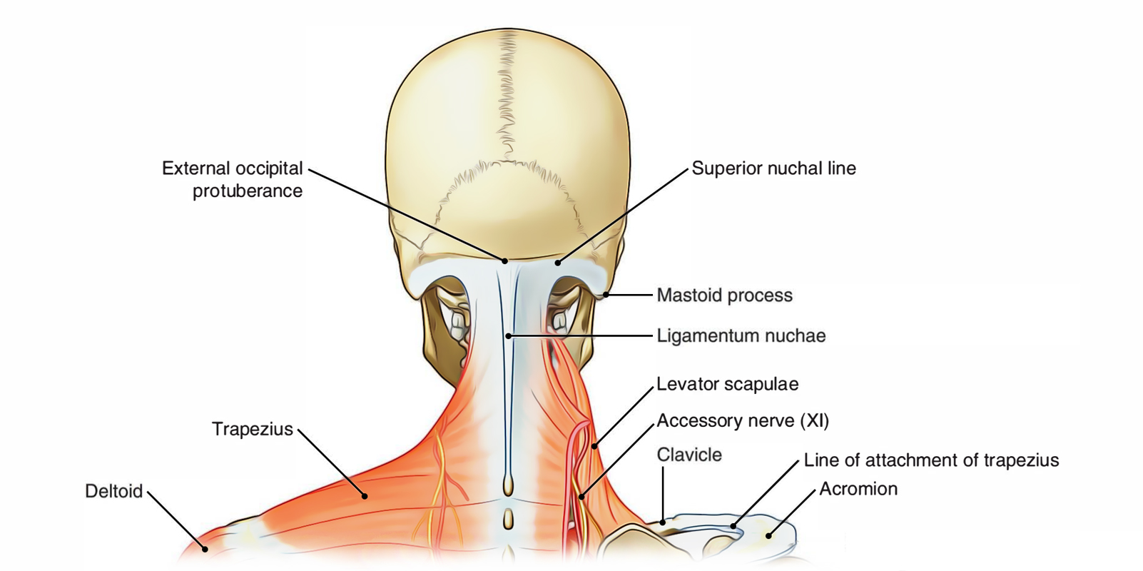
Neck: Posterior Region
The following structures can be felt in the posterior region of the neck:
- External occipital protuberance: It can be easily felt with inion at its peak in the upper end of nuchal furrow in the posterior midline of the neck.
- Superior nuchal line: It can occasionally be felt as a curved bony line with concavity below going from external occipital protuberance to the mastoid process.
- Spine of 7th cervical vertebra (vertebra bulge): It can be felt at the lower end of nuchal furrow particularly when the neck is bent.
- Ligamentum nuchae: It’s lifted when the neck is bent and stretches from spine of C7 vertebra below to the external occipital protuberance above.
Clinical Significance Medically, the posterior region of neck is very significant due to the debilitating damage it endures from whiplash injury or a busted neck.
Triangles of the Neck
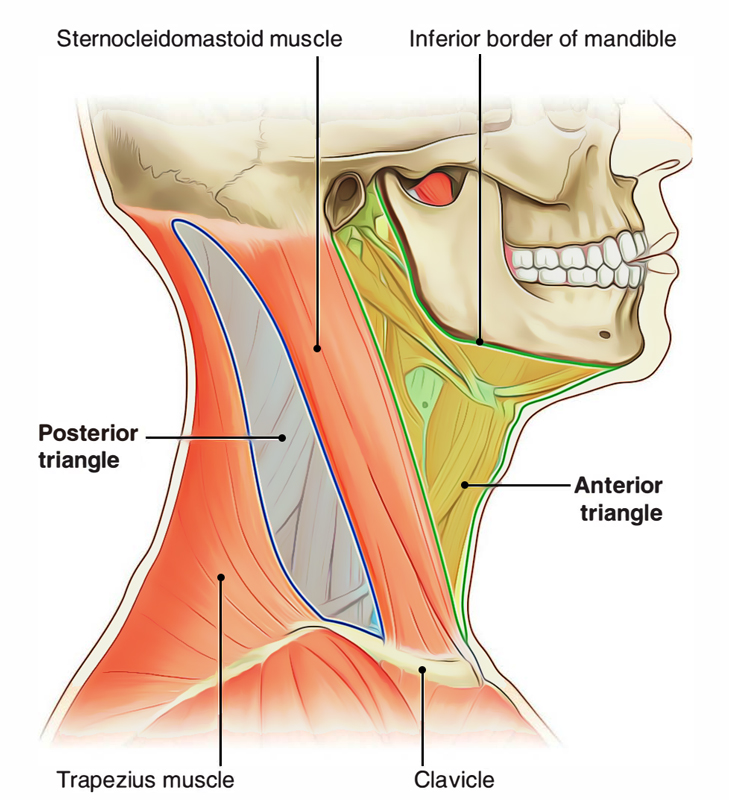
Neck: Triangles of the Neck
The neck is conventionally split into different triangles. The sternocleidomastoid muscle transects the side of neck obliquely on every side and splits it into anterior and posterior cervical triangles.

 (60 votes, average: 4.75 out of 5)