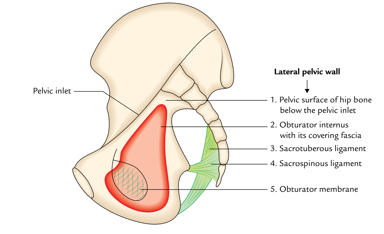
Obturator membrane is a tough, thin fibrous sheet which covers the obturator foramen of hip bone. It is superiorly perforated and creates a channel known as the obturator canal through which the obturator vessels and nerves travel across towards the medial section of the thigh.
Primarily, the fibers of the obturator membrane have transverse setting. It gives attachment to the internal and external obturator muscles. The obturator membrane is attached with the pelvic surface of the inferior ramus of ischium on its lower lateral angle. The membrane and the boundaries of the obturator foramen are connected with each other.

Obturator Membrane
Relations
- Oblique diameter at mid-pelvic cavity spreads out via the lower end of the sacroiliac joint of one side till the middle of the obturator membrane of the other side.
- The lateral wall of pelvis is created via:
- Pelvic surface of the hip bone inferior to the pelvic inlet.
- Obturator membrane
- Sacrotuberous ligaments
- Sacrospinous ligaments
- Obturator internus muscle with its covering fascia.
- Obturator Internus originates via:
- Obturator membrane
- Margins of the obturator foramen (with the exception of at the obturator canal).
- Pelvic surface of the ileum among the obturator foramen and the greater sciatic notch.
- Obturator externus arises via:
- Outer surface of the anterior half of the obturator membrane.
- Neighboring anterior and inferior boundaries of the obturator foramen.
- Anterior border of ischium creates the posterior boundary of the obturator foramen and also gives connection to the obturator membrane.
- Upper boundary of conjoint ischiopubic ramus creates the lower margin of obturator foramen as well as attaches with the obturator membrane.
- Inferior boundary of superior pubic ramus creates the upper border of obturator foramen and gives connection to the obturator membrane and spares an opening for the obturator canal.
Clinical Significance
Surgical Value
There are various surgical procedures for this region but obturator sling procedures are different. Obturator sling procedures traverse:
- Adductor muscles
- Obturator internus
- Obturator Externus muscles
Single-incision slings are dependent on the positioning of anchors via the obturator internus muscle as well as the obturator membrane.

 (54 votes, average: 4.67 out of 5)