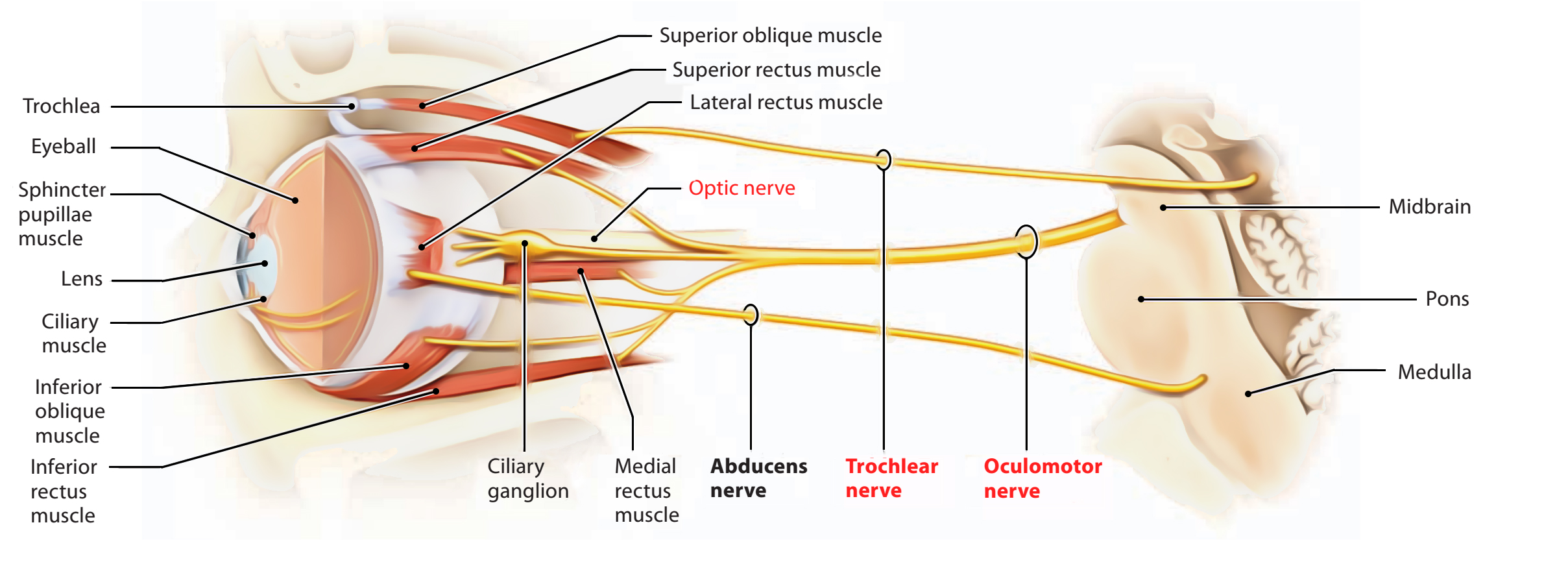
As its name indicates, it moves the eye. It supplies majority of the muscles of the eye and plays a main part in lodging of the eye. Oculomotor nerve is the 3rd cranial nerve. It’s a motor nerve only.

Oculomotor Nerve
Functional Parts and Nuclei
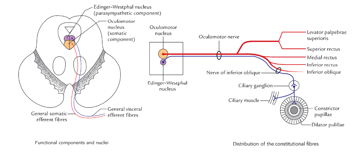
General somatic efferent fibres: All extraocular muscles with the exception of lateral rectus (supplied by6th cranial nerve) and superior oblique (supplied by 4th cranial nerve), Mnemonic: A3, LR6S04 are supplied by them. The GSE fibres originate from the somatic component of oculomotor nucleus (also termed the somatic motor nucleus).

Oculomotor Nerve: Functional Parts and Nuclei
General visceral efferent fibres: They originate from the parasympathetic component of oculomotor nucleus (also termed the Edinger Westphal nucleus). They supply the sphincter pupillae and ciliaris muscles. All these relay in the ciliary ganglion and are preganglionic parasympathetic fibres. The postganglionic parasympathetic fibres supply the sphincter pupillae and ciliaris muscles and originate from the ganglion.
Origin and Course

Oculomotor Nerve: Origin
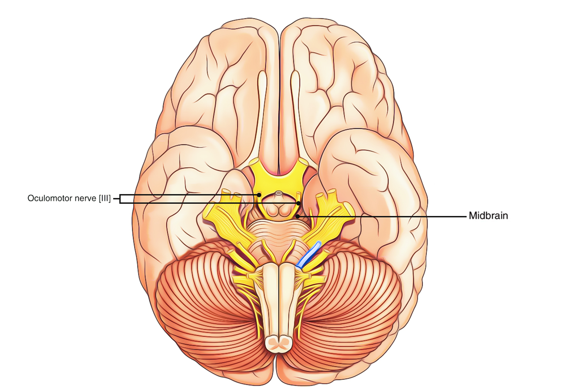
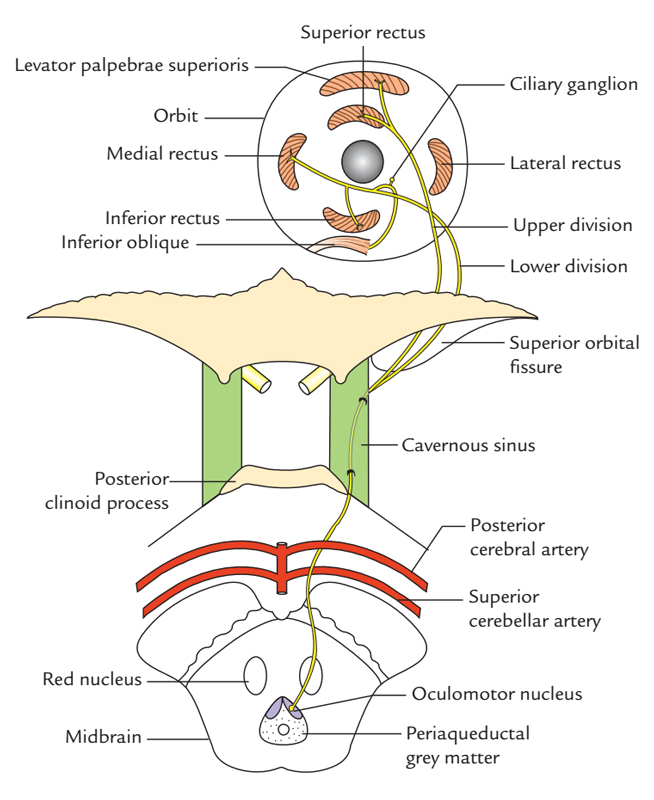
The oculomotor nerve originates from the oculomotor sulcus on the medial aspect of the cerebral peduncle of the midbrain and appears in the interpeduncular fossa. It runs forwards and laterally between the posterior cerebral and superior cerebellar arteries and lateral to the posterior communicating artery, goes through the tentorial notch of tentorium cerebelli to get to the middle cranial fossa.
Here it pierces the dura mater in the oculomotor triangle being located in between the free and connected margins of tentorium cerebelli in the roof of the cavernous sinus and enters the lateral wall of the cavernous sinus where it is located above the trochlear, ophthalmic and maxillary nerves and lateral to the internal carotid artery.
Distribution
In the anterior part of the cavernous sinus, the nerve splits into upper and lower sections.

Oculomotor Nerve: Origin, Course and Distribution
Both sections go into the orbit via the superior orbital fissure inside the common tendinous ring. The nasociliary nerve intercedes between both sections. The smaller upper section enters above the optic nerve on the inferior surface of superior rectus (which it supplies) and after that goes through the superior rectus to supply the levator palpebrae superioris.
The bigger inside section of the oculomotor nerve enters below the optic nerve and promptly supplies 3 branches which supply the medial rectus, inferior rectus and inferior oblique muscles. The nerve to inferior oblique supplies motor root (parasympathetic root) to the ciliary ganglion positioned in the posterior part of the orbit. The postganglionic fibres from using this ganglion run via short ciliary nerves and supply the sphincter pupillae and ciliaris muscles.
Clinical Significance
Lesions of Oculomotor Nerve
The damage of oculomotornerve may happen as a result of following variables:
- Compression by aneurysm of the posterior communi-cating artery as it enters between posterior cerebral and superior cerebellar arteries.
- Compression by aneurysm of the internal carotid artery as it goes through the lateral wall of the cavernous sinus.
- Compression by transtentorial uncal herniation as it goes through the tentorial notch.
The damage of oculomotor nerve medically presents as:
- Ptosis (drooping of the upper eyelid), as a result of paralysis of the levator palpebrae superioris.
- Lateral strabismus (i.e., lateral squint), because of paralysis of the medial rectus and consequent unopposed activity of the lateral rectus muscle.
- Dilated and fixed pupil, because of paralysis of the sphincter pupillae and consequent unopposed actions of the dilator pupillae.
- Loss of lodging, because of paralysis of the medial rectus, sphincter pupillae and ciliaris muscles.
- Double vision or diplopia happens on looking medially, inferiorly and superiorly, as a result of paralysis of the medial rectus, inferior rectus and inferior oblique muscles.
- Proptosis (bulge of the eyeball) because of relaxation of the muscles of the eyeball.

 (75 votes, average: 4.80 out of 5)