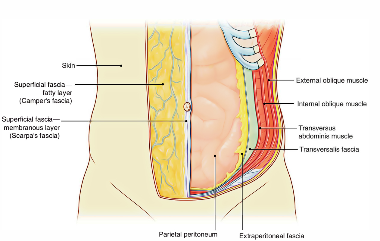
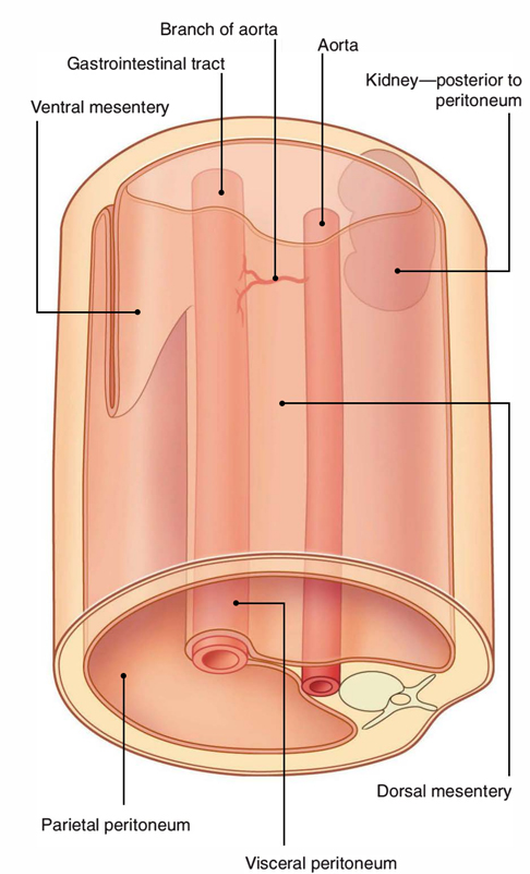
The peritoneum lines the interior of the abdominopelvic cavity and is a large slim serous membrane. Peritoneum forms the biggest serous sac of the body and is comprised of a strong layer of elastic tissue lined with the basic squamous epithelium. It resembles the pleura and serous pericardium in including parietal and visceral layers.

Peritoneum
These layers are separated from each other by a prospective space which is filled with a thin capillary film of fluid called peritoneal cavity. This assists in the movement of those parts which are confined by the visceral layer, i.e. of the abdominal viscera and fluid lubricates the two layers of the peritoneal cavity.
Layers of Peritoneum
At first the peritoneum forms a closed sac, however peritoneum splits into two layers when it ends up being invaginated by a variety of abdominal viscera:
- An outer parietal layer.
- An inner visceral layer.
Mesenteries are the folds of peritoneum through which viscera are attached. Therefore, the peritoneum provides two layers:
- Parietal layer known as parietal peritoneum.
- Visceral layer known as visceral peritoneum.

Peritoneum: Layers
The parietal peritoneum is a basic layer lining the internal surface of the abdominopelvic walls. The visceral layer has complicated plan. It forms folds, which surround the elaborately folded and securely loaded gut tube.
Differences
- The peritoneum is a closed serous sac creased with mesothelium, in the male.
- In the woman, since the peritoneum interacts with the outside through uterine tubes, uterus, and vagina, the peritoneum is not a closed sac. It is likewise lined by mesothelium as in male.
- The peritoneum covering the ovaries is lined by cuboidal epithelium.
Folds of Peritoneum
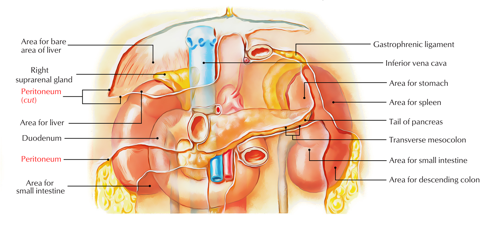
These are formed by the visceral layer of the peritoneum. The peritoneal folds suspend numerous organs within the abdomen. Within the abdominal cavity, these organs are mobile. The degree and instructions of their mobility depend upon the size and instructions of the folds. The peritoneal folds likewise give pathways for passage to nerves, vessels, and lymphatics apart from enabling mobility to organs.
The organs that lie outside the peritoneal cavity (retroperitoneal organs) are fixed and stable. Some organs have mesenteries however eventually lose their mesenteries by a procedure called zygosis and end up being retroperitoneal; they are at first suspended by the peritoneal folds.

Peritoneum: Folds
Functions
The functions of peritoneal folds are as follows:
- To give mobility to the viscera.
- To give passage to vessels and nerves.
Classification
The peritoneal folds are categorized into three types:
Mesentery/Mesocolon: The fold suspending the small intestine is called mesentery and the fold suspending the colon is called mesocolon.
- Greater omentum is a fold linking the stomach with the transverse colon.
- Lesser omentum is a fold linking the stomach with the liver.
- Gastrosplenic omentum is a fold linking the stomach with the spleen (in general use it is called gastrosplenic ligament).
Ligaments: They are the folds that link organs to the abdominal wall or to each other. The examples are:
- Gastrosplenic ligament (in between stomach and spleen).
- Lienorenal ligamentum (in between kidney and spleen).
- Coronary ligaments (in between liver and diaphragm).
Functions of Peritoneum
- For the free motion of the abdominal viscera peritoneum gives a slippery surface.
- In between the surrounding viscera peritoneum gives a moist surface to prevent friction.
- Within mesothelial lining of peritoneum it protects the viscera against infection by phagocytic cells.
- By twisting around the inflamed site and sealing the site of perforation by its fold it avoids the spread of infection, e.g: greater omentum.
- For absorption of fluids peritoneum gives a large surface area.
- Peritoneum gives recovery power for its mesothelial cells, which can change into fibroblasts.
- Peritoneum gives storage of fat in its folds, e.g., greater omentum, especially in overweight individuals.
Peritoneal Cavity
- In the human body, the peritoneal cavity is the biggest and most complicated serous sac.
- It is a prospective space in between the parietal and visceral layers of the peritoneum.
- In the male it is a closed cavity, however in the female it interacts with the outside through ostia of uterine tubes, uterus, and vagina; for that reason pelvic infections prevail in the women.
Subdivisions of the Peritoneal Cavity
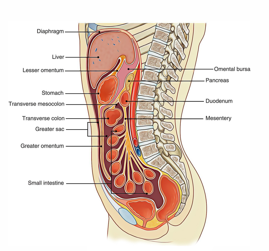
The peritoneal cavity might be broadly divided into two parts:
- Greater sac.
- Lesser sac (or omental bursa).

Peritoneum: Cavity
The greater sac is the larger (primary) compartment of the peritoneal cavity and extends throughout the entire breadth and length of the abdomen. The lesser sac is the smaller compartment of the peritoneal cavity, which lies behind the stomach, liver, and lesser omentum as a diverticulum from the greater sac. The two sacs interact with each other through the foramen epiploicum.
Greater Sac
It is the main part of the peritoneal cavity and within this sac extends all the peritoneal organs.
Supracolic and Infracolic Compartments of the Peritoneal Cavity
The abdominal part of the peritoneal cavity is divided into anterosuperior supracolic and posteroinferior infracolic compartments by transverse colon and its mesentery – the transverse mesocolon.
Supracolic Compartment
The supracolic compartment is mainly underneath the cover of costal margin and diaphragm. The accessories of the liver to the diaphragm and anterior abdominal wall specify the subdivisions of the supracolic compartment.
It lies anterior to the pancreas, duodenum, kidney, and suprarenal glands. The supracolic compartment of peritoneal cavity surrounds the liver, stomach, spleen, and the superior part of the duodenum.
Infracolic Compartment
The infracolic compartment is filled with the coils of jejunum and ileum, and enveloped by ascending, transverse, and coming down colons. By the root of the mesentery of the small intestine, the infracolic compartment underneath the level of transverse mesocolon is split into right and left infracolic areas.
It is confined on the right side by the ascending colon, above by the transverse mesocolon, and on the left side by the mesentery of the small intestine. The right infracolic space is triangular with its peak being located underneath at the ileocaecal junction.
The left infracolic space is larger than the right infracolic space and is quadrangular fit. Underneath it is constant throughout the pelvic brim in to the pelvic cavity. It is bounded above by the transverse mesocolon, on the right at the root of mesentery, and on the left by the coming down colon.


