Flattened epithelium(mesothelium) acts as a lining on a serous membrane which is similar to pleura known as peritoneum. A watery lubricant is secreted by the lining epithelium called the serous fluid.

Pleura
Layers of the Pleura
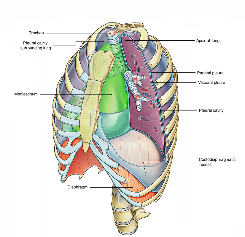
2 layers that are contained by the pleura: (a) visceral pleura (b) parietal pleura. Pleural Cavity is the name given to the wet space between both the layers.
Visceral Pleura (Pulmonary Pleura)
The visceral pleura entirely covers the top layer of the lung with the exception of at the hilum and along the connection of the pulmonary ligament. It also extends in the depths of the fissures of the lungs. It’s firmly adherent to the lung surface and can’t be divided from it.

Pleura: Layers
Parietal Pleura
The parietal pleura is thicker compared to the visceral pleura and lines the walls of the pulmonary cavity.
Subdivisions
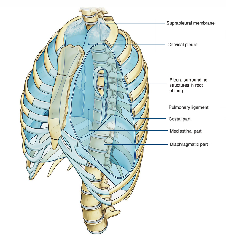
With the aim of description, it’s usual to break up parietal pleura, as stated by the surface, which it lines, covers or the region where it is located. Therefore parietal pleura is split into the following 4 parts:.
- Costal pleura.
- Diaphragmatic pleura.
- Mediastinal pleura.
- Cervical pleura.
Costal Pleura
It lines the inner surface of the thoracic wall (being composed of ribs, costal cartilages, and intercostal spaces) to which it’s loosely connected by a thin layer of loose areolar tissue termed endothoracic fascia. In living beings, endothoracic fascia is easily separable from the thoracic wall.
Diaphragmatic Pleura
It covers the superior surface of the diaphragm. In silent respiration, the costal and diaphragmatic pleura are in opposition to every other below the inferior border of the lung.
Mediastinal Pleura
It lines the corresponding outermost layer of the mediastinum and creates its lateral boundary. It’s represented as a cuff over the root of the lung and becomes constant with the visceral pleura.
Cervical Pleura
It’s the dome of parietal pleura, which extends into the root of the neck about 1 inch (2.5 cm) above the medial end of clavicle and 2 inches (5 cm) above the 1st costal cartilage. It’s termed cupola and covers the apex of the lung. Consequently, extreme care ought to be taken while penetrating this area with anesthetic needle. It’s covered by the suprapleural membrane.
Connections Of Cervical Pleura
The relationships of cervical pleura are as follows:
- Arteriorly: Subclavian artery and scalenus anterior muscle. Posteriorly: Neck of 1st rib and structures passing in front of it.
- Laterally: Scalenusmedius muscle.
- Medially: Great vessels of the neck.

 (50 votes, average: 4.68 out of 5)