The Portal Vein is a significant venous route [about 3 inches (7.5 cm) in length], which collects blood from (i) abdominal and pelvic parts of the alimentary tract (with the exception of lower part of the anal canal), (ii) gallbladder, (iii) pancreas, and (iv) spleen, and transports it to the liver.
The significant features of the portal vein are:
- It gives about 80% of the blood that flows via the liver.
- Its tributaries and branches include up to 1-third of the overall volume of blood in the whole body.
- The portal vein and its tributaries are devoid of valves.
- It carries the products of digestion of carbs, proteins, and other nutrients from the intestine and also products of red cell destruction (etc.) from the spleen to the liver.
- It splits into branches which, like those of hepatic artery, eliminate their blood into sinusoids of the liver, that are emptied by hepatic veins into the IVC.
- It starts like vein from capillary bed of the gut and ends to be an artery in the hepatic sinusoids.
Formation, Course, and Branches
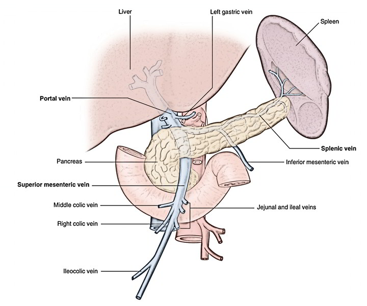
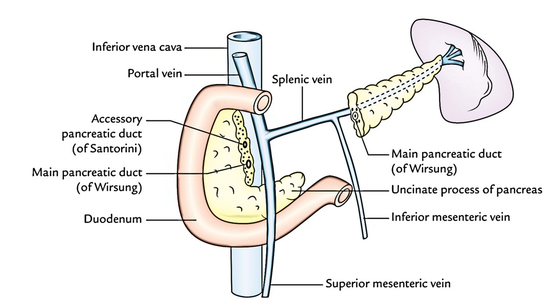
Composed behind the neck of pancreas, the portal vein is unified by superior mesenteric vein and splenic vein in the level of L2
It runs upward and a little to the right supporting the neck of pancreas, then ascends posterior to the very first part of the duodenum to goes into the right free edge of the lesser omentum. It finishes at the right end of porta hepatis by splitting into a right branch and a left branch.
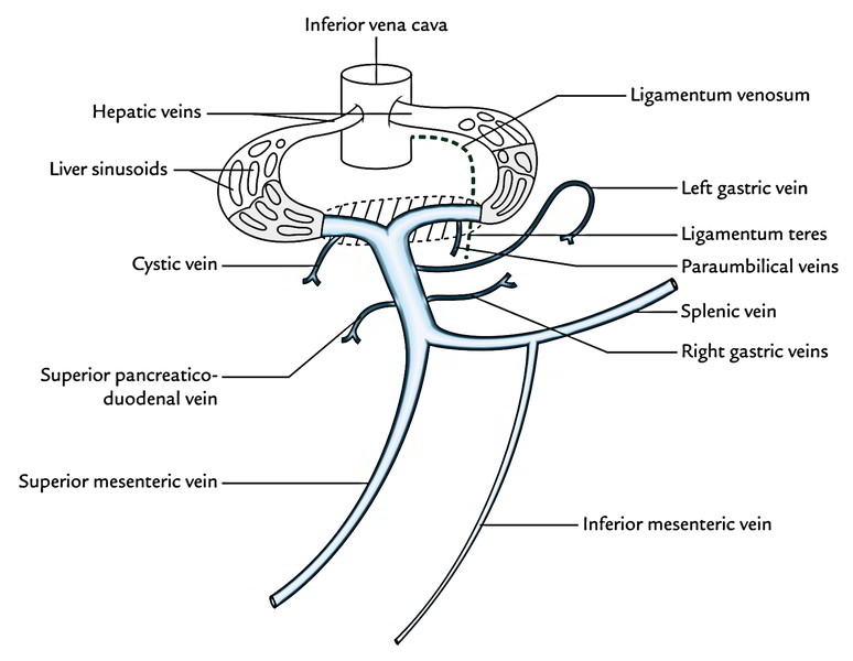
- Right branch: It’s shorter and wider and enters the right lobe of liver after receiving the cystic vein.
- Left branch: It’s longer and narrower. It enters to the left end of porta hepatis supplying branches to the caudate and quadrate lobes. Subsequently unsites with ligamentum teres and ligamentum venosum gets paraumbilical veins (which run with ligamentum teres in the falciform ligament) before entering into the left lobe of the liver.
Intrahepatic course
After going into the liver, every branch of the portal vein divides and split, like those of the hepatic artery to finish finally into the hepatic sinusoids. Here the portal venous blood blends with the hepatic arterial blood, and is divided from the liver cells by an individual layer of phagocyte and fenestrated epithelium. From hepatic sinusoids the blood is drained by hepatic veins into the IVC.
Parts and Relationships
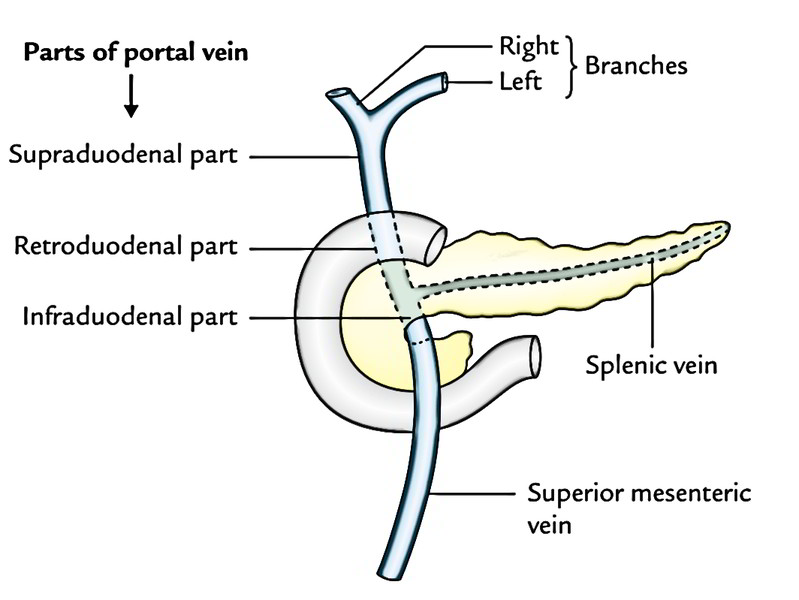
With the aim of description, the portal vein is split into 3 parts:
- Infraduodenal part which is located below the initial part of the duodenum.
- Retroduodenal part which is located posterior to the initial part of the duodenum,.
- Supraduodenal part which is located above the very first part of the duodenum in the right free margin of lesser omentum.
The relationships of distinct parts of the portal vein are as follows:
Infraduodenal Part
- Anterior: Neck of pancreas.
- Posterior: IVC.
Retroduodenal Part
- Anterior: First part of the duodenum, Bile duct, Gastroduodenal artery.
- Posterior: IVC.
Supraduodenal Part
- Anterior: Hepatic artery (on the left) and bile duct (on the right).
- Posterior: IVC.
Tributaries
The portal vein gets the subsequent tributaries:
- Splenic vein, a bigger formative tributary.
- Superior mesenteric vein, a smaller formative tributary.
- Superior pancreaticoduodenal veinjoins the portal vein behind the very first part of duodenum.
- Right and left gastric veinsjoin the portal vein the right free margin of lesser omentum.
- The left gastric veinat the cardiac end of the stomach gets a couple of esophageal veins from the lower end of esophagus.
- The right gastric veinreceives the prepyloric vein (of Mayo) which runs vertically in front of the pylorus.
- Cystic veinjoins the right branch of the portal vein before it enters the right lobe of the liver.
- Paraumbilical veinsare small veins that run along the ligamentum teres in the falciform ligament and join the left branch of the portal vein before it enters the left lobe of the liver.
Portocaval (Portosystemic) Anastomoses
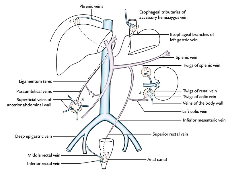
There are various ssites in the abdominal cavity where anastomosis exists between the portal and systemic venous systems. These communications between the veins of the portal vein and caval (systemic) systems create essential routes of collateral circulation in cases of portal vein obstruction.
The essential ssites of portocaval anastomoses are as follows:
- Umbilicus: Here the left branch of the portal vein interacts with all the superficial veins of the anterior abdominal wall around umbilicus, via paraumbilical veins (of Sappey). In portal vein obstruction, the superficial veins around the umbilicus become distended and tortuous (varicosity). This whorl of notable distended tortuous (snakelike) veins around the umbilicus is called caput medusae1- an indication of diagnostic value to the clinicians.
- Lower end of esophagus: Here the esophageal tributaries of left gastric vein (emptying into the portal vein) anastomose with esophageal tributaries of accessory hemiazygos vein (systemic).
- In portal vein obstruction as in liver cirrhosis, these collateral channels become distended and tortuous, creating esophageal varices, which might rupture causing hematemesis (vomiting of blood) and might even bleed to death.
- Anal canal: Here the superior rectal (hemorrhoidal) vein which finally empties into the portal vein veinanastomoses with middle and inferior rectal veins, the tributaries of internal iliac (systemic) vein. The distension and dilatation of these anastomotic channels result in the formation of hemorrhoids or piles which might be liable for recurrent bleeding per annum.
- Extraperitoneal surfaces of retroperitoneal organs. Veins of retroperitoneal organs like duodenum, ascending colon, and descending colon (portal vein) anastomose with the retroperitoneal veins of the posterior abdominal wall and renal capsule (systemic). The renal vein anastomosis with splenic and azygos veins.
- Naked area of liver: Here the hepatic venules (portal vein) anastomose with phrenic and intercostal (systemic) veins.
Caput head of Medusa (in Greek Medusae). Medusa (a legendary woman) was the daughter of Phorcys and captivated Neptune with her gold hair and became by him the mother of Pegasus. As a punishment, Minerva altered her hair into serpents and empowered her eyes so that everything her eyes looked upon transformed to stone. Perseus slew and decapitated her and from the blood that dropped serpents sprung.
Anastomoses Between The Portal and Systemic Venous Systems
Site of anastomosis | Veins forming portocaval anastomosis | Clinical signs |
|---|---|---|
| Lower third of the esophagus | Left gastric vein U Esophageal veins draining into azygos vein | Esophageal varices |
| Umbilicus | Paraumbilical veins U Superficial veins of anterior abdominal wall | Caput medusae |
| Mid-anal canal | Superior rectal vein U Middle and inferior rectal veins | Hemorrhoids |
Clinical Significance
Portal hypertension: Obstruction of the portal vein or its branches leads to the rise in the portal venous pressure named portal hypertension, i.e., pressure above 40 mmHg (normal being 5-15 mmHg). This contributes to enlargement of collateral channels. The most ordinary cause of portal hypertension is alcoholic cirrhosis of the liver.
The impacts of portal hypertension are:
- Splenomegaly (enlargement of the spleen).
- Ascsites (build-up of fluid in the peritoneal cavity).
- Esophageal varices.
- Hemorrhoids.

 (64 votes, average: 4.53 out of 5)