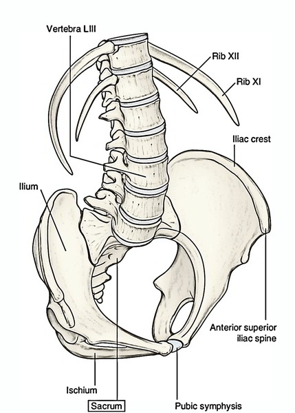
Sacrum is termed as a large flattened triangular/ wedge shaped bone created by the fusion of 5 sacral vertebrae. The sacrum articulates on either side with all the hip bone to create the sacroiliac joint. Sacrum has the following functions:
- Sacrum Creates posterior part of the bony pelvis.
- Supports the vertebral column.
- Conducts the weight of the body to the pelvic girdle via the sacroiliac joints.

Sacrum
Anatomical Position
To keep the sacrum in anatomical position, hold it in this manner that:
- Smooth pelvic surface of sacrum faces downward and forward and its rough dorsal surface faces upward and backwards.
- Upper surface of the body of the very first sacral vertebra pitches forward at an angle of about 30 °
- Upper end of the sacral canal is directed nearly upward and somewhat backwards.
In an articulated skeleton, the sacrum is put obliquely like a wedge between both hip bones. When lumbar vertebral column is in position, an angle is composed between L5 and L1 vertebrae. This angle is named sacrovertebral/lumbosacral angle. It measures about 210 ° and is being rounded by the intervening disk that is considerably thicker in front than behind.
General Features
The sacrum presents the following general features:
- Base
- Apex
- 4 surfaces: pelvic, dorsal, and 2 lateral (left and right) surfaces
- Sacral canal

Sacrum: Features
Base
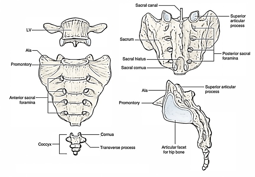
The base of the sacrum is directed upward and forward. It’s created by the superior surface of the very first sacral vertebra. The sacrum of the base is split into 3 parts: median part and right and left lateral parts. The median part is composed by the oval upper surface of the body of the very first sacral vertebra. Its anterior border is referred to as sacral promontory. The left and right lateral parts are fan shaped and represent the upper surfaces of lateral masses of sacrum referred to as alae. The lateral masses represent the fused costal and transverse aspects of the sacral vertebrae.
- Upper surface of the body of the very first sacral vertebra articulates with the fifth lumbar vertebra.
- Vertebral foramen being located behind the body is triangular and leads into the sacral canal.
- Pedicles are short and directed backwards and laterally.
- Laminae are rather oblique.
- Transverse processes are immense and fused with the corresponding costal transverse component of lower sacral vertebrae to create the broad sloping lateral masses named alae of sacrum.
- The spinous process creates the first spinous tubercle.
- The superior articular processes are notable and project upward. They flank the superior opening of sacral canal.
Apex
It’s the lower narrow dull extremity created by the inferior outermost layer of the body of the fifth sacral vertebra. It bears an oval facet for articulation with all the coccyx to create the sacrococcygeal joint.
Surfaces
Pelvic Surface
The pelvic surface of sacrum is concave and directed downward and forward. It presents these features:
- 4 transverse ridges that suggest the lines of fusion of 5 sacral vertebrae.
- Lateral to transverse ridges are 4 anterior sacral foramina via which ventral rami of upper 4 sacral nerves come out.
- The part of bone lateral to foramina is the lateral mass.
Dorsal Surface
The dorsal surface of sacrum is rough, uneven, and convex.
- In the median plane, there’s median sacral crest that bears 3 or 4 spinous tubercles representing spinous processes of fused sacral vertebrae.
- Lateral to median crest, the posterior surface is composed by fused laminae of upper 4 sacral vertebrae.
- The laminae of the fifth sacral vertebra don’t join behind, creating a U shaped gap referred to as sacral hiatus.
- Lateral to laminae are 4 articular tubercles in accordance together with the superior articular process of the very first sacral vertebra.
- The inferior articular processes of the fifth sacral vertebra project downward as sacral cornua on either side of sacral hiatus.
The most notable features on the dorsal surface of sacrum is presence of 5 vertical crests:
- Median sacral crest, created by the fusion of sacral spines.
- 2 intermediate sacral crests, 1 on either side of median crest, are created by the fusion of articular processes of sacral vertebrae.
- 2 lateral sacral crests, 1 on every side of lateral to intermedial crest, are created by the fusion of transverse processes of sacral vertebrae.
- Sacral hiatus: A U-shaped gap at the lower end.
Lateral Surfaces
Every lateral surface is the lateral aspect of lateral mass formed by the fusion of transverse and costal components of sacral vertebrae.
- It’s wide above and narrow below.
- Lower narrow part turns unexpectedly medially to create the inferolateral angle of the sacrum.
- Upper wider part bears an inverted L shaped auricular surface anteriorly and a deeply pitted area posteriorly. The auricular surface is so called due to its similarity to the auricle of the external ear. It goes on to the upper 3 or 3 and a half sacral vertebrae.
Sacral Canal
- The sacral canal is composed by the superimposed vertebral foramina of the sacral vertebrae.
- The upper end of the canal is directed upward in line with vertebral canal of lumbar vertebrae and its lower end opens at the sacral hiatus.
- The sacral canal interacts with all the pelvic and dorsal sacral foramina via the intervertebral foramina.
Contents of Sacral Canal
- Lower part of cauda equina (sacral and coccygeal nerve roots).
- Filum terminale.
- Spinal meninges.
- Lateral sacral vessels.
The dural tube and arachnoid mater go up to the 2nd sacral vertebra.
Structures Coming Via Sacral Hiatus
- Fifth sacral (S5) nerves.
- Coccygeal (Cx1) nerves.
- Filum terminale.
Clinical Significance
Sacralization
The term sacralization means incorporation of the fifth lumbar (L5) or first coccygeal vertebra (C1) in the sacrum. In sacralization the number of sacral foramina is raised unilaterally or bilaterally.
Lumbarization
In this state, the very first sacral (S1) vertebra is divided from the sacrum and fused with the fifth lumbar vertebra (L5). Consequently, the number of sacral foramina is reduced to 3 pairs.
Special Features (Attachments and Relations)
Base
- The upper surface of the body of the very first sacral vertebra articulates with the L5 vertebra to create the lumbosacral joint.
- The smooth medial part of the ala of sacrum is associated with the subsequent 4 structures from medial to lateral side: 1. Sympathetic chain. 2. Lumbosacral trunk. 3. Iliolumbar artery. 4. Obturator nerve.
- The ventral ramus of L5 nerve is really tight that it grooves the ala. The rough lateral part of the ala provides origin to iliacus muscle anteriorly and connection to the lumbosacral ligament posteriorly.
Apex
It bears an oval facet, which articulates with the body of first coccygeal vertebra to create the sacrococcygeal joint.
Surfaces
Pelvic Surface
It presents the following specific features:
- The median sacral vessels are associated with the pelvic surface in the median plane.
- The sympathetic trunks are linked to the pelvic surface along the medial margins of pelvic sacral foramina.
- The upper 2 -and-half sacral vertebrae are associated with the parietal peritoneum with the exception of the site of connection of the medial limb of the sigmoid mesocolon.
- The lower 2 -and-half vertebrae are linked to the rectum, but split from S3 vertebra by bifurcation of the superior rectal artery.
- The piriformis muscle originates from the bodies of middle 3 sacral vertebrae in an E-shaped style.
- The pelvic sacral foramina carry ventral rami of the corresponding upper 4 sacral nerves and a branch from lateral sacral arteries.
Dorsal Surface
It presents the following unique features:
- The 4 dorsal foramina conduct the dorsal rami of the corresponding upper 4 sacral spinal nerves.
- The erector spinae muscle originates from an elongated U shaped linear area affecting the constant rows of the spinous and transverse tubercles.
- The multifidus originates from the area enclosed by U-shaped origin of erector spinae. The dorsal rami of upper 4 sacral spinal nerves get to the surface by piercing multifidus and erector spinae muscles.
Lateral Surfaces
The auricular surface/facet articulates together with the corresponding auricular outermost layer of the ilium to create sacroiliac joint.
- The rough area behind the auricular surface provides connection to strong interosseous sacroiliac ligament.
- The lower narrow part of the lateral surface opposite the inferolateral angle provides connection to 4 structures. From behind forwards these are: a) Gluteus maximus b) Sacrotuberous ligament c) Sacrospinous ligament d) Coccygeus
- The final structure (i.e., coccygeus) encroaches on the pelvic surface of the sacrum.
- Sacral index: It’s the ratio between the length and width of the sacrum and is expressed by the formula, width x 100/length.
- The sacral index is higher in females than males. The typical sacral index in a female is 116 and in a male it’s 112.
Differences Between the Male and Female Sacrum
Features | Male | Female |
|---|---|---|
| Base of sacrumSacral index | Width of articular area (body of S1 vertebra)is more than the length of ala of one side, i.e., body is broad and alae are narrowLess (sacrum is relatively longer and narrower) | Width of articular area (body of S1 vertebra) is either equal or less than the length of ala of one side, i.e., body is narrow and alae are broadMore (sacrum is relatively shorter and broader) |
| Pelvic surface | 1. Smoothly curved, “C” shaped (curvature of the pelvic surface is gradual from above downward) | 1. Abruptly curved, “J” shaped (lower part of the pelvic surface abruptly curves forward, curvature being most marked between S1 and S2 segments and between S3 and S5 segments) |
| 2. Concavity of sacrum is shallower | 2. Concavity of sacrum is deeper | |
| Auricularsurface | 1. Extends on to the upper 3 or 3% of sacral vertebrae | 1. Extends on to the upper 2 or 2% of the sacral vertebrae |
| 2. Relatively larger and less obliquely set | 2. Relatively smaller and more obliquely set | |
| 3. Concavity of dorsal border of the auricular surface is less marked | 3. Concavity of dorsal border of the auricular surface is more marked |
Ossification
The ossification of sacrum isn’t of much clinical interest. Nonetheless, people should just understand that since the sacrum is a composite bone created by the fusion of 5 sacral vertebrae, it ossifies by multiple centers.
Test Your Knowledge
Sacrum

 (53 votes, average: 4.89 out of 5)