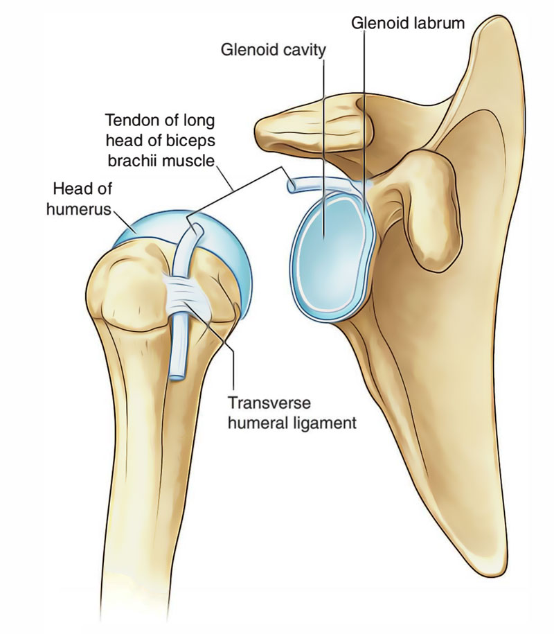
The Glenohumeral Joint (Shoulder joint) is a synovial ball and socket articulation between the head of the humerus and the glenoid cavity of the scapula. Glenohumeral joint is multiaxial with a broad range of movements provided in the cost of skeletal stability. Shoulder joint stability is provided instead, by the rotator cuff muscles, the long head of the biceps brachii muscle, related bony processes, and extracapsular ligaments. Movements at the shoulder joint contain flexion, extension, abduction, adduction, medial rotation, lateral rotation, and circumduction. The articular surfaces of the glenohumeral joint are the large spherical head of the humerus and the small glenoid cavity of the scapula. Every of the surfaces is covered by hyaline cartilage.

Glenohumeral Joint
The glenoid cavity is deepened and expanded peripherally by a fibrocartilaginous collar (the glenoid labrum), which connects to the margin of the fossa. Superiorly, this labrum is continuous with the tendon of the long head of the biceps brachii muscle, which connects to the supragle- noid tubercle and goes through the articular cavity superior to the head of the humerus.
The shoulder joint is known to be the least stable among other joints but also the most movable joint of the body. The students must thoroughly study the shoulder joint as it usually undergoes recurrent dislocations and is the most common joint to dislocate. Shoulder Joint is also known as Glenohumeral joint.
Type
The shoulder joint is a ball-and-socket type of synovial joint.
Articular Surfaces
The shoulder joint is formed by articulation of large round head of humerus with the relatively shallow glenoid cavity of the scapula. The glenoid cavity is deepened slightly but effectively by the fibrocartilaginous ring called glenoid labrum.
Ligaments
The ligaments of the glenohumeral joint are as follows:
1. Capsular ligament (joint capsule): The thin fibrous layer of the joint capsule covers the glenohumeral joint. It is attached medially to the margins of the glenoid cavity past the glenoid labrum and laterally to the anatomical neck of the humerus, except inferiorly where it extends downwards 1.5 cm or more on the surgical neck of the humerus. Medially the attachment extends beyond the supraglenoid tubercle thus wrapping the long head of biceps brachii within the joint cavity.
The synovial membrane lines the inner surface of the joint capsule and reflects from it to the glenoid labrum and humerus regarding the articular margin of the head. The synovial cavity of the glenohumeral joint shows the following features:
- It forms tubular sheath around the tendon of biceps brachii where it is located in the bicipital groove of the humerus.
- It communicates with subscapular and infraspinatus bursae, around the joint.
Thus there are three apertures in the joint capsule:
- An opening between the tubercles of the humerus for the passage of tendon of long head of biceps brachii.
- An opening located anteriorly inferior to the coracoid process to allow interaction between the synovial cavity and subscapular bursa.
- An opening located posteriorly to allow communication between synovial cavity and infraspinatus bursa.
2. Glenohumeral ligaments: There are three thickenings in the anterior part of the fibrous capsule; to strengthen it. These are called superior, middle, and inferior glenohumeral ligaments. They are visible only from interior of the joint.
A defect exists between superior and middle glenohumeral ligaments, which obtain importance in the anterior dislocation of the shoulder joint.
3. Coracohumeral ligament: It is a strong band of fibrous tissue that passes from the base of the coracoid process to the anterior part of the greater tubercle of the humerus.
4. Transverse humeral ligament: It is a broad fibrous band, which links the bicipital groove between the greater and lesser tubercles. This ligament changes the groove into a canal that provides passage to the tendon of long head of biceps surrounded by a synovial sheath.
Accessory Ligaments
The accessory ligaments of the shoulder joint are as follows:
- Coracoacromial ligament: It extends between coracoid and acromion processes. It protects the superior component of the joint.
- Coracoacromial arch: The coracoacromial arch is formed by coracoid process, acromion process, and coracoacromial ligament between them. This osseoligamentous structure forms a protective arch for the head of humerus above and prevents its superior displacement above the glenoid cavity. The supraspinatus muscle passes under this arch and exists deep to the deltoid where its tendon mixes with the joint capsule. The large subacromial bursa lies between the arch superiorly and tendon of supraspinatus and better tubercle of humerus inferiorly. This enhances the movement of supraspinatus tendon.
Bursae Related to the Shoulder Joint
Several bursae relate to the shoulder joint but the important ones are as follows :
- Subscapular bursa: It lies between the tendon of subscapularis and the neck of the scapula; and protects the tendon from friction from the neck. This bursa normally communicates with the joint cavity of glenohumeral joint.
- Subacromial bursa: It lies between the coracoacromial ligament and acromion process above, and supraspinatus tendon and joint capsule below. It continues downwards beneath the deltoid, hence it is sometimes also described as subdeltoid bursa.
It is the biggest synovial bursa in the body and helps with the movements of supraspinatus tendon under the coracoacromial arch. - Infraspinatus bursa: It lies between the tendon of infraspinatus and posterolateral aspect of the joint capsule. It may sometime communicate with the joint cavity.
The bursae around the shoulder joint are clinically vital as some of them interact with synovial cavity of the fhoulder joint. Hence, opening a bursa may mean entering into the cavity of the glenohumeral joint.
Relations Of the Shoulder Joint
The shoulder joint is related:
Superiorly: to coracoacromial arch, subacromial bursa, supraspinatus muscle, and deltoid muscle.
Inferiorly: to long head of triceps, axillary nerve and posterior circumflex humeral vessels.
Anteriorly: to subscapularis, subscapular bursa, coraco- brachialis, short head of biceps brachii, and deltoid.
Posteriorl: to infraspinatus, teres minor, and deltoid.
Clinical Correlation
A portion of epiphyseal line of proximal humerus is intracapsular, thus, septic arthritis of the shoulder joint may occur following metaphyseal osteomyelitis.

 (63 votes, average: 4.59 out of 5)