A spinal nerve is composed of many nerves, which carries motor, sensory, and autonomic signals between the spinalcord and the body. Human body consist of 31 pairs of spinal nerves, one on each side of the vertebral column.
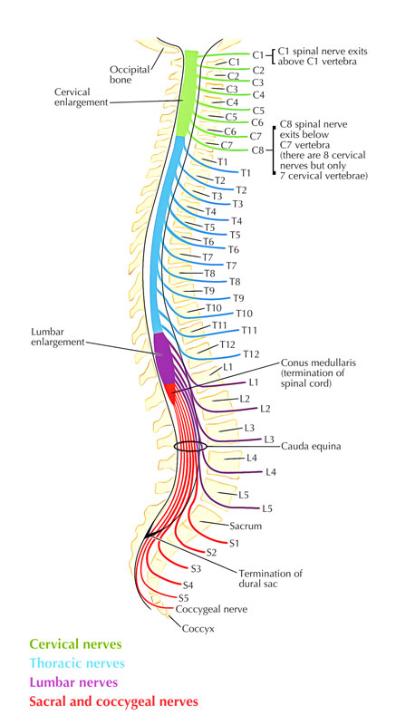
The 31 sets of spinal nerves are segmental in circulation and arise via the vertebral canal in between the stalks of surrounding vertebrae. There are:
- 8 sets of cervical nerves (Cl to C8)
- 12 thoracic (T1 to T12)
- 5 lumbar (LI to L5)
- 5 sacral (SI to S5)
- 1 coccygeal (Co)
Each nerve is connected to the spinal cord by a posterior root and an anterior root. After leaving the vertebral canal, every spinal nerve sections into:
- A posterior ramus: jointly, the small posterior rami innervate the back.
- An anterior ramus: the much greater anterior rami innervate most other areas of the body other than the head, which is innervated primarily, however not exclusively, by cranial nerves.
Primarily significant visceral parts of the PNS (sympathetic trunk and prevertebral plexus) of the body are likewise connected with the anterior rami of spinal nerves. The anterior rami create the significant somatic plexuses (cervical. brachial, lumbar, and sacral) of the body.

Spinal Nerves
Intervertebral Formina and Spinal Nerves
The foramen is created in between surrounding vertebral arches and is carefully related to intervertebral joints. Each spinal nerve leaves the vertebral canal laterally via an intervertebral foramen:
- The superior and inferior margins are created by notches in surrounding stalks.
- The posterior margin is created by the articular processes of the vertebral arches and the associated joint.
- The anterior border is created by the intervertebral disc in between the vertebral bodies of the surrounding vertebrae.
Any pathology that occludes or decreases the size of an intervertebral foramen, such as bone loss, herniation of the intervertebral disc, or dislocation of the zygapophysial joint (the joint in between the articular procedures), can impact the function of the associated spinal nerve.
Innervation of the Back
Posterior branches of spinal nerves innervate the intrinsic muscles of the back and surrounding skin. The cutaneous distribution of these posterior rami extends into the gluteal region of the lower limb and the posterior element of the head.
Structure
Each spinal nerve includes a dorsal root (containing afferent fibers) and a ventral root (consisting of afferent fibers). The dorsal root is missing in the first cervical and coccygeal nerves. The common spinal nerve trunk is gotten in touch with the sympathetic chain of paravertebral ganglia that lie on either side of the vertebral column via the white and gray rami. The dorsal and ventral roots take a trip a short range, permeate the dura, and enter into the intervertebral foramen, within the dural sac surrounding the spinal cord. The spinal ganglion (dorsal root ganglion) lies within the intervertebral foramen and consists of the neurons that produce afferent fibers entering into the spinal cord. The dorsal and ventral roots create the common spinal nerve trunk and are signed up with distal to the spinal ganglion.
Plexuses
- Axons via the anterior rami of spinal nerves, except for thoracic nerves T2-T12, do not go straight to the body structures they supply.
- Instead, by accompanying numerous varieties of axons via anterior rami of surrounding nerves they create systems on both the left and right sides of the body.
- Such a system of axons is called a plexus (PLEK-sus = braid or system).
- The cervical plexus, brachial plexus, lumbar plexus, and sacral plexus are the primary plexuses.
- Emerging via the plexuses are nerves bearing names that are frequently descriptive of the basic areas they serve or the course they take. A smaller coccygeal plexus is likewise present.
- Each of the nerves in turn might have various branches called for the certain structures they innervate.
Cervical Plexus
There are eight cervical nerves (C1 to C8). C1 to C7 emerges via the vertebral canal over their particular vertebrae and C8 emerges in between vertebrae CVII and TI.
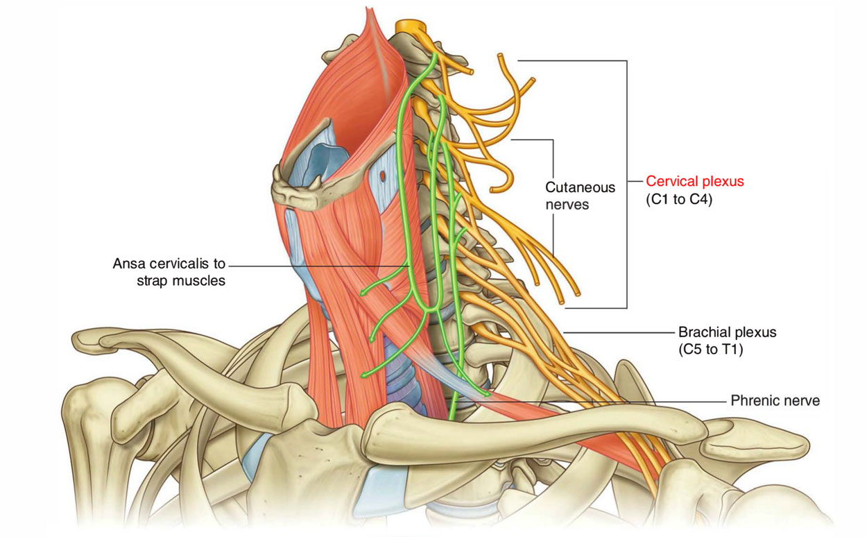
The anterior rami of C1 to C4 create the cervical plexus. The significant branches via this plexus supply:
- The strap muscles.
- The diaphragm (phrenic nerve).
- Skin on the anterior.
- Lateral parts of the neck.
- Skin on the upper anterior thoracic wall.
- Skin on the inferior parts of the head.
The anterior rami of C5 to C8 form the brachial plexus, which innervates the upper limb, along with a large part of the anterior ramus of Tl.
Thoracic Plexus
T1-12, reinforce the muscles that lie in between the ribs (intercostal muscles). These likewise assist breathing by pulling the chest outwards and upwards, pulling the lungs in the same direction. The lungs broaden, helping them fill with air.
Primary breathing muscles are the diaphragm and the intercostal muscles. Lower thoracic spinal nerves T6-12 supply power to abdominal muscles. These muscles assist to cough and expel matter via airway. Abdominal muscles are likewise crucial in balance and posture.
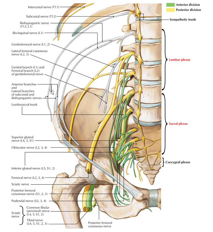
Lumbar and Sacral Plexus
Nerves in the lumbar (L) and sacral (S) areas of spinal cord power leg muscles for walking, running and jumping.

Lumbar and Sacral Plexus
- L2 powers muscles that flex or bend hip joint.
- L3 powers quadriceps muscle at the knee so the leg straighten up.
- L4 powers muscles enabling ankle to flex and draw the foot back to head (dorsi-flexion) around ankle.
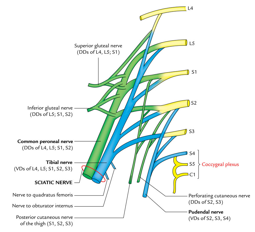
- S1 powers muscles enabling it to flex and foot and toes to point downwards (plantar flexion) around ankle
- S2-S4 powers external sphincter muscles of anal canal and urethra
Coccygeal Plexus

Coccygeal Plexus
The coccygeal plexus stems from the S4, S5, and C01 spinal nerves. It is adjoined with the lower part of the sacral plexus. The only nerve in this plexus is the anococcygeal nerve, which serves sensory innervation of the skin in the coccygeal region.


 (49 votes, average: 4.80 out of 5)