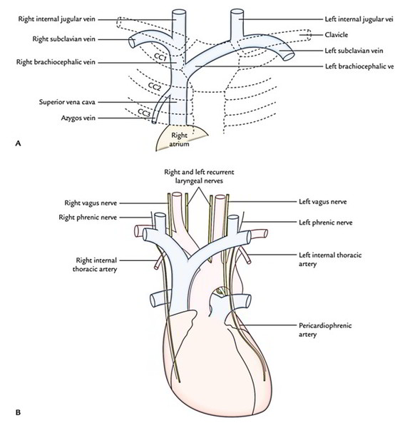
Superior Vena Cava is about 7 cm long and 1.25 cm in diameter. Its location is in the superior and middle mediastina. The extrapericardial part is located in the superior mediastinum and intrapericardial part is located in the middle mediastinum. It collects deoxygenated blood from the upper half of the body (i.e., head and neck, upper limbs, thoracic wall, and upper abdomen) and drains it into the right atrium. Different collateral pathways develop depending on the site of obstruction.The signs of obstruction of superior vena cava appear first in mediastinal syndrome.
A-Formation, Course and Termination B-Relations.
Relations
Anterior:
A. Right internal thoracic vessels.
B. Margin of right lung and pleura.
C. Chest wall.
Posterior:
A. Trachea (posteromedial).
B. Right pulmonary artery and right bronchus.
To The Left:
A. Ascending aorta (anteromedial).
B. Brachiocephalic artery.
To The Right:
A. Right phrenic nerve and pericardiophrenic vessels.
B. Right lung and pleura.
Formation, Course, And Termination
The superior vena cava is composed at the lower border of the right 1st costal cartilage by the union of left and right brachiocephalic (innominate) veins. It enters vertically downwards behind the right border of the sternum and pierces the pericardium in the level of the right 2nd costal cartilage, and opens/ends into the upper part of the right atrium at the lower border of the right 3rd costal cartilage. It has no valves in its lumen because gravity facilitates the blood flow in it.
Subdivisions
The superior vena cava is subdivided into the following 2 parts:
A. Extrapericardial part (in superior mediastinum).
B. Intrapericardial part (in middle mediastinum).
Tributaries
A. Left and right brachiocephalic veins.
B. Azygos vein, which arches over the root of the right lung and opens into SVC just before it pierces fibrous pericardium.
C. Mediastinal and pericardial veins.
Brachiocephalic Veins
There are 2 brachiocephalic veins: (a) right and (b) left. Every of them is composed behind the sternoclavicular joint by the union of corresponding internal jugular and subclavian veins. They unite to create SVC. Both are devoid of valves.
Differences Between Left And Right Brachiocephalic Veins
Right brachiocephalic vein Left brachiocephalic vein Length • Short (2.5 cm) • Long (6 cm) Course • Vertical (runs vertically downwards from right sternoclavicular joint to the lower margin of the right 1st costal cartilage) • Oblique (runs obliquely across the superior mediastinum from left sternoclavicular joint to the lower margin of the right 1st costal cartilage) Tributaries • Right vertebral vein • Left vertebral vein • Right internal thoracic vein • Left internal thoracic vein • Right inferior thyroid vein • Left inferior thyroid vein • First right posterior intercostal vein •First left posterior intercostal vein
Clinical Significance
Obstruction Of SVC And Development Of Collateral Pathways
The SVC could be obstructed (compressed) at 2 sites: (a) above the opening of azygos vein (i.e., in superior mediastinum), and (b) below the opening of azygos vein (i.e., in the middle mediastinum).
If SVC is obstructed above the opening of azygos vein, the venous blood from the upper half of the body is shunted to right atrium via azygos vein. The main collateral pathways are provided by the superior intercostal veins. The superficial veins of chest wall don’t receive sufficient blood to cause their prominence. If at all they become notable, the prominence is limited up to the costal margin only.
If SVC is obstructed below the opening of the azygos vein, the venous blood from the upper half of the body is returned to the right atrium via inferior vena cava via the collateral pathways, created between the tributaries of superior and inferior vena cavae (caval- caval shunt). Medically in this condition, a subcutaneous anastomotic channel between the superficial epigastric vein and lateral thoracic vein (thoraco-epigastric vein) is observed on the anterior aspect of the thoraco-abdominal wall.


 (47 votes, average: 4.78 out of 5)