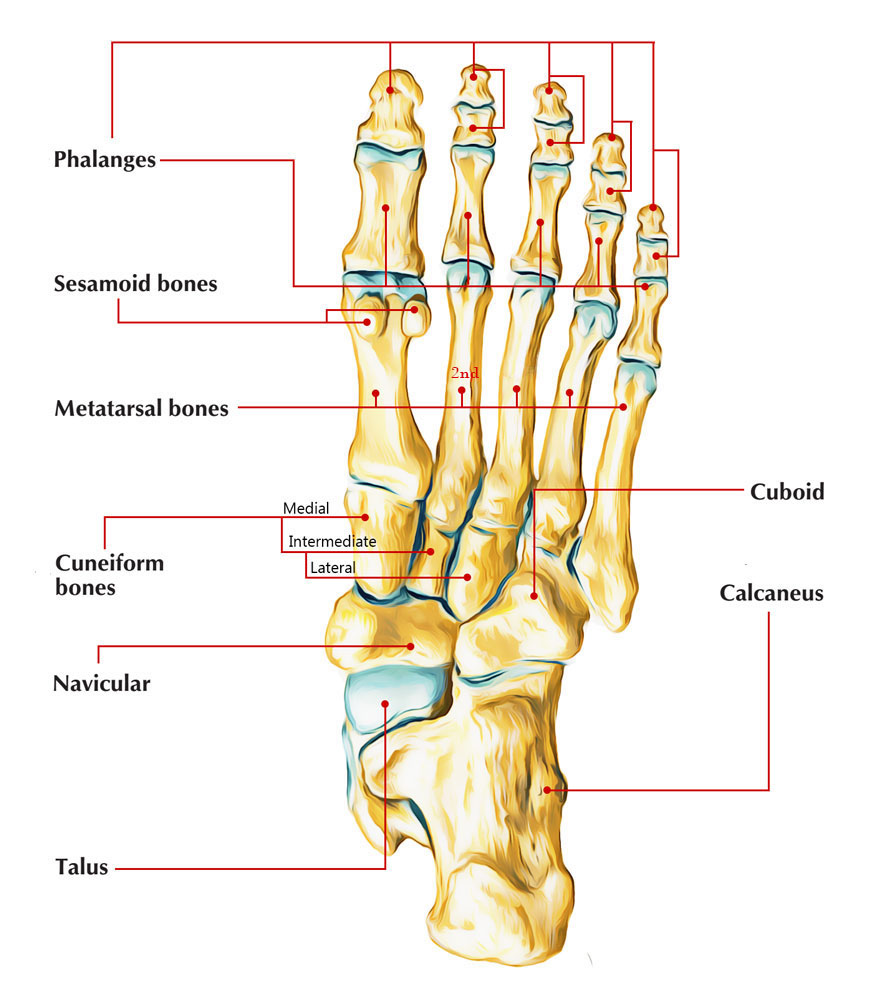
Structure
The second metatarsal bone is the longest of the metatarsal bones, being extended backwards into the hollow created by the three cuneiform bones. Its base is wide superiorly, slim and unequal inferiorly.

The Second Metatarsal
Side
- It gives four articular sides: one behind, of a triangular kind, for joining with the second cuneiform; one at the upper part of its medial side, for joining along with the first cuneiform; as well as two on its lateral side, an upper as well as lower, divided by an unequal non-articular interval.
- A fifth aspect sometimes exists for joining with the first metatarsal; it is elliptical in shape, and is located on the medial side of the body near the base.
- All of these lateral articular sides is split into two by an vertical rim; the two anterior aspects integrate by the third metatarsal; the two posterior along with the third cuneiform.
Clinical Significance
Injuries
- Separated fractures of the second metatarsal are unusual. Fracture of this particular bone was associated with fractures of other metatarsals in fractures of the lesser second via fifth metatarsals.
- The most common system of injury was indirect, such as leaping from a height of less than 5 feet.
- Other head-on systems consisted of items such as a rock, table, or chair falling on the foot.
- The fractures were seldom displaced, other than with extreme trauma. The majority of these fractures can be treated with below-knee walking casts as soon as the swelling is down.
Tension Fracture
- The second metatarsal is more frequently based on tension fracture. The injury takes place early in the second years, is typically called a March fracture, and also generally takes place in runners.
- The injury has actually seen in inactive kids who unexpectedly increase their walking or running activity.
- The child experiences consistent pain under the metatarsal arch.
- The preliminary radiographs might be unfavorable; however a bone scan is positive.
- Periosteal cortical hypertrophy or brand-new bone normally exists within 2 weeks after problems of pain. Therapy frequently includes using a hard-soled shoe; seldom is a cast suggested. Displacement of the pieces is seldom seen.
- The second metatarsal is the longest and also most strictly fixed metatarsal. Repeated trauma to the articular side of the distal end of the second metatarsal might trigger this injury.
- The injury generally takes place after extensive training for running sports and has actually been seen in passionate young soccer gamers. Conventional therapy by rest or using a below-knee walking cast is advised.
- Small osteochondral pieces might be left in the second metatarsophalangeal joint. Open debridement might be needed to prevent degenerative joint disease.
Balter – Harris Type IV Fractures
The injury is seldom dislocated, liven fractures along with plantar flexion have the tendency to renovate. The condylar fractures are generally oblique Balter-Harris type IV fractures and seldom lead to development injuries. Fractures of the chondroepiphysis of the second metatarsal take place most often as Salter – Harris type II fractures of the neck.
x

 (47 votes, average: 4.63 out of 5)