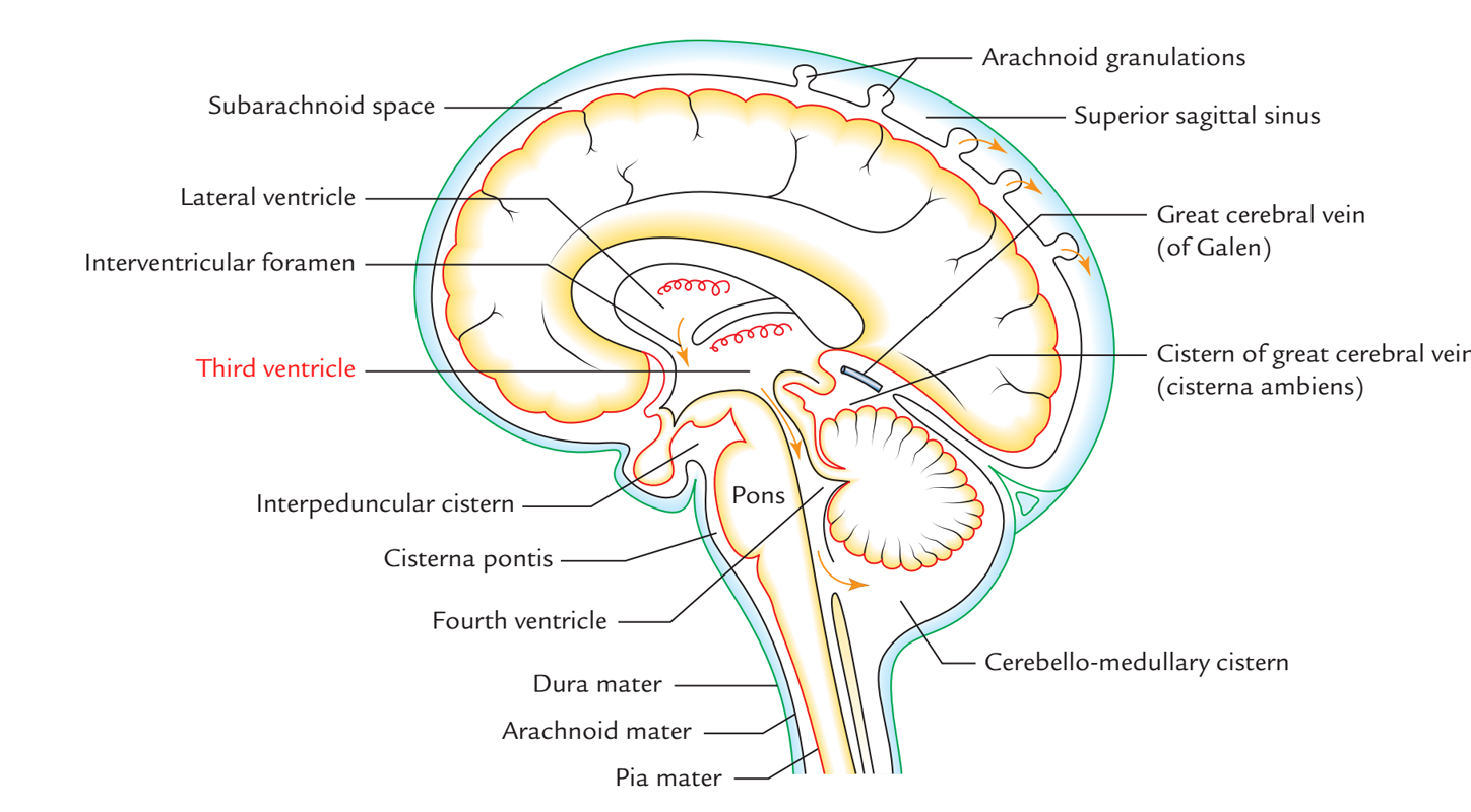
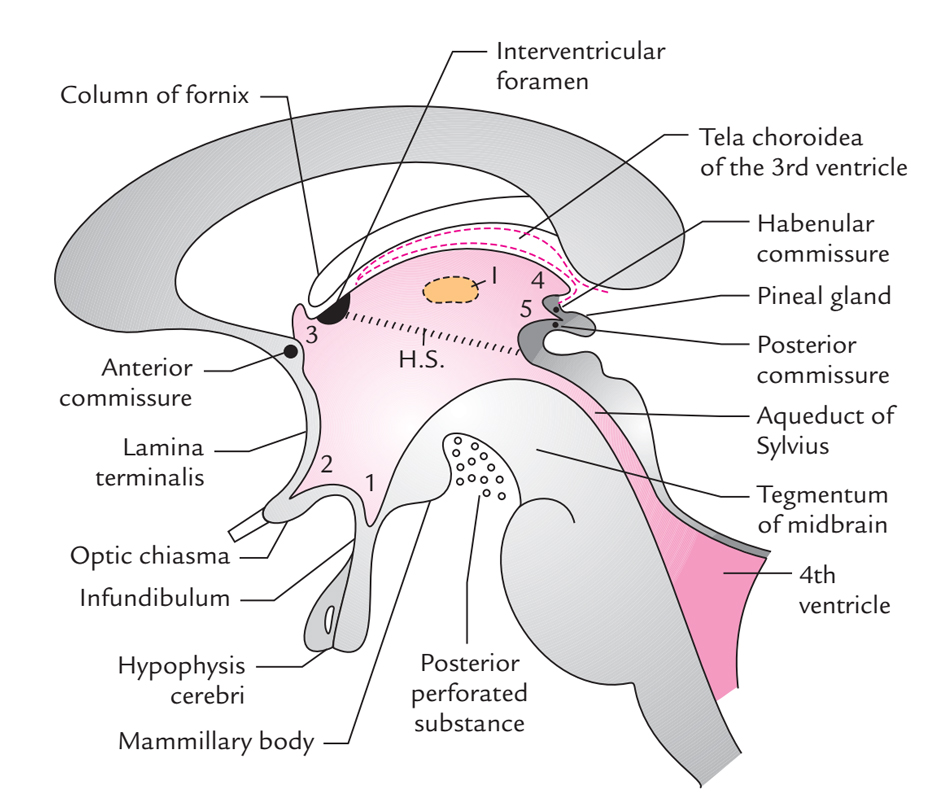
The 3rd ventricle is the cavity of diencephalon. It’s a midline slit-like cavity situated between 2 thalami and part of hypothalamus. It goes from the lamina terminalis anteriorly to the superior end of the cerebral aqueduct of the midbrain posteriorly. The cavity of the 3rd ventricle is lined by ciliated columnar epithelium, the ependyma and traversed horizontally by a mass of grey matter, the interthalamic adhesion, joining the 2 thalami. The outline of the cavity is atypical because of the presence of several diverticula or recesses.

Third Ventricle
Anteriorly on every side, the 3rd ventricle interacts with the whole lateral ventricle via interventricular foramen (of Monro) and posteriorly with the 4th ventricle via cerebral aqueduct (of Sylvius).
Bounds
The 3rd ventricle has anterior wall, posterior wall, roof, floor and 2 lateral walls.

Third Ventricle: Boundaries
Anterior wall is composed from above downward by:
- Anterior column of fornix
- Anterior commissure
- Lamina terminalis
Posterior wall: created from above downward by:
- Pineal gland
- Posterior commissure
- Commencement of cerebral aqueduct.
Roof: Created by the ependyma that stretches across the upper limits of 2 thalami.
Floor: Created from before backwards by:
- Optic chiasma
- Tuber cinereum and infundibulum
- Mammillary bodies
- Posterior perforated substance
- Tegmentum of the midbrain
All structures of the floor belong to interpeduncular fossa with the exception of the optic chiasma and tegmentum of the midbrain.
Lateral wall:
- Marked by a curved sulcus, the hypothalamic sulcus going from the interventricular foramen to the upper end of the cerebral aqueduct. The sulcus divides the lateral wall into a bigger upper part and a smaller lower part.
- The bigger upper part of the lateral wall is composed by the medial surface of the anterior two-third of the thalamus.
- The smaller lower part of the lateral wall is composed by the hypothalamus and it’s constant with the ventricular floor.
- The 2 lateral walls of the 3rd ventricle are normally closely approximated, for this reason in coronal section of the brain the cavity of the 3rd ventricle appears as a median vertical slit.
Recesses
The cavity of the 3rd ventricle extends into the surrounding structures as pocket-like protrusions referred to as recesses. All these are as follows:

Third Ventricle: Recesses
- Infundibular recess: It’s a deep tunnel-shaped recess extending downward via the tuber cinereum into the infundibulum, i.e., the stalk of the pituitary gland
- Optic (or chiasmatic) recess: It’s an angular recess situated in the junction of the anterior wall and the floor of the ventricle just above the optic chiasma.
- Anterior recess (vulva of the ventricle): It’s a triangular recess which extends anteriorly in front of interventricu-lar foramen and behind anterior commissure between the diverging anterior columns of the fornix.
- Suprapineal recess: It’s a reasonably capacious blind diverticulum, which goes posteriorly above the stalk of the pineal gland and below the tela choroidea.
- Pineal recess: It’s a small diverticulum which widens posteriorly between the superior and inferior laminae of the stalk of the pineal gland
Choroid Plexus and Tela Choroidea
The tela choroidea in the roof of the 3rd ventricle is triangular in shape. The choroid plexus of the 3rd ventricle hangs downward from the tela choroidea as 2 longitudinal anteroposterior vascular peripheries.
Clinical Significance
Impediment of Third Ventricle
The 3rd ventricle being a narrow slit-like space is easily obstructed by localized brain tumors or congenital defects. The obstacle results in excessive accumulation of CSF inside the brain, leading to an increased intracranial pressure in adults and in hydrocephalus in youngsters.

 (60 votes, average: 4.62 out of 5)