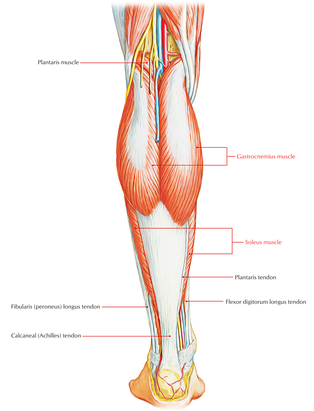
The triceps surae muscles are a muscle of the calf, providing the calf volume on the rear side. It expands via the femur to the calcaneus, from behind the knee and the tibia. It is comprised of the gastrocnemius muscle, made up of medial together with lateral packages, as well as the soleus muscle. The triceps surae muscle gives a propulsion movement which allows us to stroll, supports the placement of the leg and raises the heel.

Triceps Surae
Anatomy and Supply
The triceps surae muscle is a three-headed muscle at the dorsal lower leg. Its heads as well as body identify the surface anatomy of the calf. It is located in the superficial posterior compartment. Its attachment, the Achilles tendon, is quickly noticeable as well as perceptible at the heel. The triceps surae muscle is supplied by the tibial nerve. It is comprised by:
- Soleus muscle: Emerges at the upper fibula, tibia as well as the tendinous arch of soleus which extends in between the tibia as well as fibula.
- Gastrocnemius muscle (left): It has its origin on the medial epicondyle (medial head) along with lateral epicondyle (lateral head) of the femur. It runs over the soleus muscle nearly completely
- The neurovascular bundle originating from the hollow of the knee (posterior tibial artery along with vein and tibial nerve) travel under the tendinous arch of soleus, afterwards that it routes in between the superficial and deep flexor muscles.
- At the distal third of the lower leg both of the soleus and gastrocnemius muscles combine in a common tendon, the achilles tendon (tendo calcaneus), which enter at the posterior calcaneus.
Attachements
- The medial head is connected to a lengthen roughening on the posterior element of the distal femur simply just behind the adductor tubercle and also above the articular surface of the medial condyle.
- Via an unique side on the upper lateral surface of the lateral femoral condyle the lateral head emerges, the place where it signs up with the lateral supracondylar line.
- At the knee, the dealing with margins of the two heads of the gastrocnemius create the lateral and medial borders of the lower end of the popliteal fossa.
- The heads of the gastrocnemius integrate to create a single lengthen muscle belly, that makes much of the soft tissue bulge determined as the calf in the upper leg.
- The muscle fibers of the gastrocnemius connect along with those of the much deeper soleus muscle to create the calcaneal tendon, which connects to the calcaneus (heel) of the foot within the lower leg.
Actions
- It is the primary plantar flexor of the foot at the ankle when the knee is expanded.
- It is also a flexor of the knee.
- It offers quick movements of the foot throughout running and leaping.
Function
- Operational activities consist of mostly activity in the sagittal plane, stabilizing throughout mobility (walking, running) as well as power leaping.
- Tightening of the triceps surae cause plantar flexion (sagittal plane) as well as stabilizing of the ankle system in the transverse plane.
Clinical Significance
Injury and Symptoms
A muscle strain injury at one or possibly more of the muscles of the triceps surae symbolizes the tear of a great deal of muscle fibers, triggered by an extreme tightening of the muscle in a violent or fast way.
There will also be existence of swelling as well as bruising and in some cases an audible pop or breeze will be heard throughout the incident of the injury. Crucial weak point in the calf may also happen, and it might be hard to stroll or stand on the toes. Symptoms of a muscle tear at the triceps surae muscle group consist of an abrupt acute pain felt at the calf, which exists even at rest.
Risk Factors
- A pre-existing absence of felxibility of the muscles at the triceps surae.
- An extreme training routine needing an essential or abrupt tightening of the calf muscles, specifically when there is absence or absence of a proper warm-up ahead of time.
- An absence of muscle strength or essential muscle tiredness.
- An improperly recovered or unhealed previous injury, such as a muscle strain.
Physiotherapy Procedures
- It is essential to keep in mind that a medical follow-up will be needed in combination to the physiotherapy procedures in order to identify the requirement of anti-inflammatory medications or of a medical surgery.
- Your therapist will also utilize a broad selection of manual treatment and myofascial release strategies, and will recommend proper extending and reinforcing workouts.
- The physiotherapy procedures will intend to decrease the pain and swelling found with using electrotherapy (such as ultrasound and TENS), ice as well as proper education concerning the activities which might aggravate condition and therefore which needs to be prevented for the time being.

 (58 votes, average: 4.81 out of 5)